1H0D

Crystal structure of Human Angiogenin in complex with Fab fragment of its monoclonal antibody mAb 26-2F


Experimental Data Snapshot

  • Method: X-RAY DIFFRACTION
  • Resolution: 2.00 Å
  • R-Value Free: 0.272 
  • R-Value Work: 0.232 
  • R-Value Observed: 0.232 

wwPDB Validation   3D Report Full Report


This is version 2.1 of the entry. See complete history


Literature

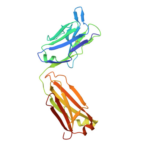
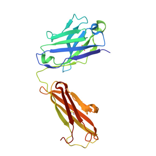
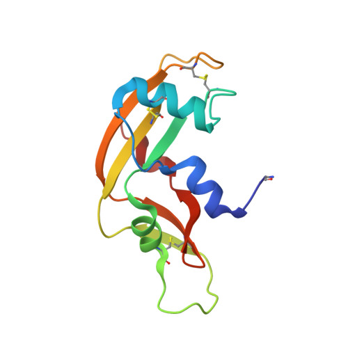
The Crystal Structure of Human Angiogenin in Complex with an Antitumor Neutralizing Antibody

Chavali, G.B.Papageorgiou, A.C.Olson, K.Fett, J.Hu, G.Shapiro, R.Acharya, K.R.

(2003) Structure 11: 875

  • DOI: https://doi.org/10.1016/s0969-2126(03)00131-x
  • Primary Citation of Related Structures:  
    1H0D

  • PubMed Abstract: 

    The murine monoclonal antibody 26-2F neutralizes the angiogenic and ribonucleolytic activities of human angiogenin (ANG) and is highly effective in preventing the establishment and metastatic dissemination of human tumors in athymic mice. Here we report a 2.0 A resolution crystal structure for the complex of ANG with the Fab fragment of 26-2F that reveals the detailed interactions between ANG and the complementarity-determining regions (CDRs) of the antibody. Surprisingly, Fab binding induces a dramatic conformational change in the cell binding region of ANG at the opposite end of the molecule from the combining site; crosslinking experiments indicate that this rearrangement also occurs in solution. The ANG-Fab complex structure should be invaluable for designing maximally humanized versions of 26-2F for potential clinical use.


  • Organizational Affiliation

    Department of Biology and Biochemistry, University of Bath, Claverton Down, BA2 7AY, Bath, United Kingdom.


Macromolecules
Find similar proteins by:  (by identity cutoff)  |  3D Structure
Entity ID: 1
MoleculeChains Sequence LengthOrganismDetailsImage
ANTIBODY FAB FRAGMENT, LIGHT CHAIN216Homo sapiensMutation(s): 0 
Entity Groups  
Sequence Clusters30% Identity50% Identity70% Identity90% Identity95% Identity100% Identity
Sequence Annotations
Expand
  • Reference Sequence
Find similar proteins by:  (by identity cutoff)  |  3D Structure
Entity ID: 2
MoleculeChains Sequence LengthOrganismDetailsImage
ANTIBODY FAB FRAGMENT, HEAVY CHAIN223Homo sapiensMutation(s): 0 
Entity Groups  
Sequence Clusters30% Identity50% Identity70% Identity90% Identity95% Identity100% Identity
Sequence Annotations
Expand
  • Reference Sequence
Find similar proteins by:  (by identity cutoff)  |  3D Structure
Entity ID: 3
MoleculeChains Sequence LengthOrganismDetailsImage
ANGIOGENIN123Homo sapiensMutation(s): 0 
UniProt & NIH Common Fund Data Resources
Find proteins for P03950 (Homo sapiens)
Explore P03950 
Go to UniProtKB:  P03950
PHAROS:  P03950
GTEx:  ENSG00000214274 
Entity Groups  
Sequence Clusters30% Identity50% Identity70% Identity90% Identity95% Identity100% Identity
UniProt GroupP03950
Sequence Annotations
Expand
  • Reference Sequence
Small Molecules
Ligands 2 Unique
IDChains Name / Formula / InChI Key2D Diagram3D Interactions
SO4
Query on SO4

Download Ideal Coordinates CCD File 
D [auth A]
I [auth B]
L [auth C]
M [auth C]
N [auth C]
D [auth A],
I [auth B],
L [auth C],
M [auth C],
N [auth C],
O [auth C],
P [auth C],
Q [auth C]
SULFATE ION
O4 S
QAOWNCQODCNURD-UHFFFAOYSA-L
GOL
Query on GOL

Download Ideal Coordinates CCD File 
E [auth A]
F [auth A]
G [auth A]
H [auth A]
J [auth B]
E [auth A],
F [auth A],
G [auth A],
H [auth A],
J [auth B],
K [auth B]
GLYCEROL
C3 H8 O3
PEDCQBHIVMGVHV-UHFFFAOYSA-N
Modified Residues  1 Unique
IDChains TypeFormula2D DiagramParent
PCA
Query on PCA
C
L-PEPTIDE LINKINGC5 H7 N O3GLN
Experimental Data & Validation

Experimental Data

  • Method: X-RAY DIFFRACTION
  • Resolution: 2.00 Å
  • R-Value Free: 0.272 
  • R-Value Work: 0.232 
  • R-Value Observed: 0.232 
  • Space Group: C 1 2 1
Unit Cell:
Length ( Å )Angle ( ˚ )
a = 120.58α = 90
b = 72.53β = 112.46
c = 86.99γ = 90
Software Package:
Software NamePurpose
CNSrefinement
DENZOdata reduction
SCALEPACKdata scaling
AMoREphasing

Structure Validation

View Full Validation Report


Entry History 

Deposition Data

Revision History  (Full details and data files)

  • Version 1.0: 2003-06-19
    Type: Initial release
  • Version 1.1: 2011-10-12
    Changes: Derived calculations, Non-polymer description, Other, Refinement description, Structure summary, Version format compliance
  • Version 2.0: 2020-03-11
    Changes: Derived calculations, Polymer sequence
  • Version 2.1: 2023-12-13
    Changes: Data collection, Database references, Derived calculations, Refinement description