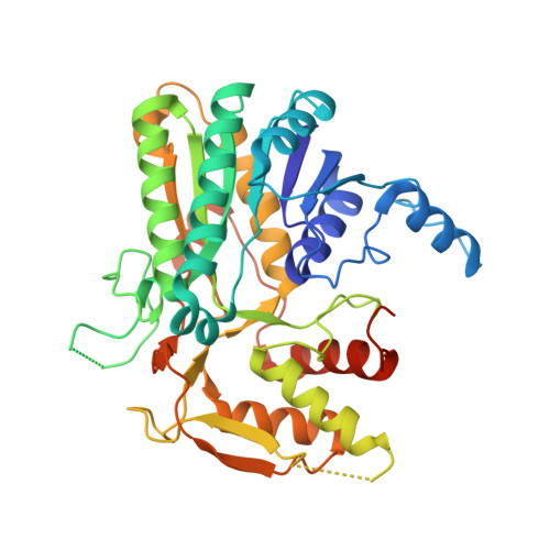
1GY8

Trypanosoma brucei UDP-galactose 4' epimerase


Experimental Data Snapshot

  • Method: X-RAY DIFFRACTION
  • Resolution: 2.00 Å
  • R-Value Free: 0.245 
  • R-Value Work: 0.216 
  • R-Value Observed: 0.216 

wwPDB Validation   3D Report Full Report


Ligand Structure Quality Assessment 


This is version 1.3 of the entry. See complete history


Literature

High-Resolution Crystal Structure of Trypanosoma Brucei Udp-Galactose 4'-Epimerase: A Potential Target for Structure-Based Development of Novel Trypanocides

Shaw, M.P.Bond, C.S.Roper, J.R.Gourley, D.G.Ferguson, M.A.J.Hunter, W.N.

(2003) Mol Biochem Parasitol 126: 173

  • DOI: https://doi.org/10.1016/s0166-6851(02)00243-8
  • Primary Citation of Related Structures:  
    1GY8

  • PubMed Abstract: 

    The crystal structure of UDP-galactose 4'-epimerase from the protozoan parasite Trypanosoma brucei in complex with the cofactor NAD(+) and a fragment of the substrates, UDP, has been determined at 2.0 A resolution (1 A = 0.1 nm). This enzyme, recently proven to be essential for this pathogenic parasite, shares 33% sequence identity with the corresponding enzyme in the human host. Structural comparisons indicate that many of the protein-ligand interactions are conserved between the two enzymes. However, in the UDP-binding pocket there is a non-conservative substitution from Gly237 in the human enzyme to Cys266 in the T. brucei enzyme. Such a significant difference could be exploited by the structure-based design of selective inhibitors using the structure of the trypanosomatid enzyme as a template.


  • Organizational Affiliation

    Division of Biological Chemistry and Molecular Microbiology, School of Life Sciences, University of Dundee, Dundee DD1 5EH, UK.


Macromolecules
Find similar proteins by:  (by identity cutoff)  |  3D Structure
Entity ID: 1
MoleculeChains Sequence LengthOrganismDetailsImage
UDP-GALACTOSE 4-EPIMERASE
A, B, C, D
397Trypanosoma bruceiMutation(s): 0 
EC: 5.1.3.2
UniProt
Find proteins for Q8T8E9 (Trypanosoma brucei)
Explore Q8T8E9 
Go to UniProtKB:  Q8T8E9
Entity Groups  
Sequence Clusters30% Identity50% Identity70% Identity90% Identity95% Identity100% Identity
UniProt GroupQ8T8E9
Sequence Annotations
Expand
  • Reference Sequence
Experimental Data & Validation

Experimental Data

  • Method: X-RAY DIFFRACTION
  • Resolution: 2.00 Å
  • R-Value Free: 0.245 
  • R-Value Work: 0.216 
  • R-Value Observed: 0.216 
  • Space Group: P 21 21 21
Unit Cell:
Length ( Å )Angle ( ˚ )
a = 102.185α = 90
b = 112.533β = 90
c = 160.591γ = 90
Software Package:
Software NamePurpose
CNSrefinement
DENZOdata reduction
SCALEPACKdata scaling
AMoREphasing

Structure Validation

View Full Validation Report


Ligand Structure Quality Assessment 


Entry History 

Deposition Data

Revision History  (Full details and data files)

  • Version 1.0: 2003-05-22
    Type: Initial release
  • Version 1.1: 2011-05-08
    Changes: Version format compliance
  • Version 1.2: 2011-07-13
    Changes: Version format compliance
  • Version 1.3: 2023-12-13
    Changes: Data collection, Database references, Other, Refinement description