1GWF

Compound II structure of Micrococcus Lysodeikticus catalase


Experimental Data Snapshot

  • Method: X-RAY DIFFRACTION
  • Resolution: 1.96 Å
  • R-Value Free: 0.190 
  • R-Value Work: 0.141 
  • R-Value Observed: 0.089 

wwPDB Validation   3D Report Full Report


Ligand Structure Quality Assessment 


This is version 1.2 of the entry. See complete history


Literature

The Structures of Micrococcus Lysodeikticus Catalase, its Ferryl Intermediate (Compound II) and Nadph Complex.

Murshudov, G.N.Grebenko, A.I.Brannigan, J.A.Antson, A.A.Barynin, V.V.Dodson, G.G.Dauter, Z.Wilson, K.S.Melik-Adamyan, W.R.

(2002) Acta Crystallogr D Biol Crystallogr 58: 1972-1982

  • DOI: https://doi.org/10.1107/s0907444902016566
  • Primary Citation of Related Structures:  
    1GWE, 1GWF, 1GWH

  • PubMed Abstract: 

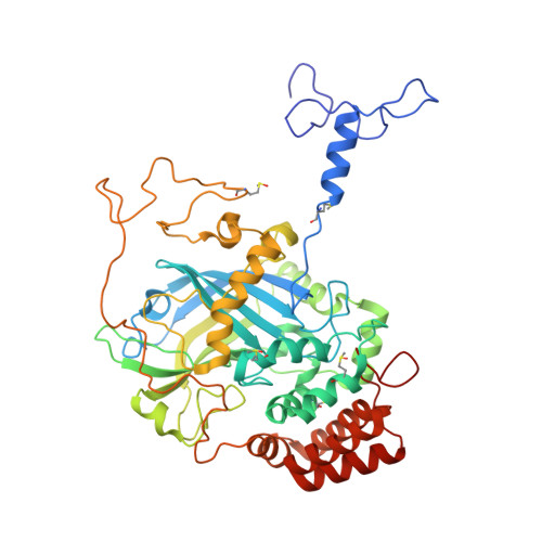
    The crystal structure of the bacterial catalase from Micrococcus lysodeikticus has been refined using the gene-derived sequence both at 0.88 A resolution using data recorded at 110 K and at 1.5 A resolution with room-temperature data. The atomic resolution structure has been refined with individual anisotropic atomic thermal parameters. This has revealed the geometry of the haem and surrounding protein, including many of the H atoms, with unprecedented accuracy and has characterized functionally important hydrogen-bond interactions in the active site. The positions of the H atoms are consistent with the enzymatic mechanism previously suggested for beef liver catalase. The structure reveals that a 25 A long channel leading to the haem is filled by partially occupied water molecules, suggesting an inherent facile access to the active site. In addition, the structures of the ferryl intermediate of the catalase, the so-called compound II, at 1.96 A resolution and the catalase complex with NADPH at 1.83 A resolution have been determined. Comparison of compound II and the resting state of the enzyme shows that the binding of the O atom to the iron (bond length 1.87 A) is associated with increased haem bending and is accompanied by a distal movement of the iron and the side chain of the proximal tyrosine. Finally, the structure of the NADPH complex shows that the cofactor is bound to the molecule in an equivalent position to that found in beef liver catalase, but that only the adenine part of NADPH is visible in the present structure.


  • Organizational Affiliation

    Structural Biology Laboratory, Department of Chemistry, University of York, York YO10 5DD, England. garib@ysbl.york.ac.uk


Macromolecules
Find similar proteins by:  (by identity cutoff)  |  3D Structure
Entity ID: 1
MoleculeChains Sequence LengthOrganismDetailsImage
Catalase503Micrococcus luteusMutation(s): 0 
Gene Names: katA
EC: 1.11.1.6
UniProt
Find proteins for P29422 (Micrococcus luteus)
Explore P29422 
Go to UniProtKB:  P29422
Entity Groups  
Sequence Clusters30% Identity50% Identity70% Identity90% Identity95% Identity100% Identity
UniProt GroupP29422
Sequence Annotations
Expand
  • Reference Sequence
Small Molecules
Ligands 4 Unique
IDChains Name / Formula / InChI Key2D Diagram3D Interactions
HEM
Query on HEM

Download Ideal Coordinates CCD File 
B [auth A]PROTOPORPHYRIN IX CONTAINING FE
C34 H32 Fe N4 O4
KABFMIBPWCXCRK-RGGAHWMASA-L
SO4
Query on SO4

Download Ideal Coordinates CCD File 
C [auth A],
F [auth A],
G [auth A]
SULFATE ION
O4 S
QAOWNCQODCNURD-UHFFFAOYSA-L
ACT
Query on ACT

Download Ideal Coordinates CCD File 
D [auth A],
E [auth A]
ACETATE ION
C2 H3 O2
QTBSBXVTEAMEQO-UHFFFAOYSA-M
O
Query on O

Download Ideal Coordinates CCD File 
H [auth A]OXYGEN ATOM
O
XLYOFNOQVPJJNP-UHFFFAOYSA-N
Modified Residues  1 Unique
IDChains TypeFormula2D DiagramParent
SME
Query on SME
A
L-PEPTIDE LINKINGC5 H11 N O3 SMET
Experimental Data & Validation

Experimental Data

  • Method: X-RAY DIFFRACTION
  • Resolution: 1.96 Å
  • R-Value Free: 0.190 
  • R-Value Work: 0.141 
  • R-Value Observed: 0.089 
  • Space Group: P 42 21 2
Unit Cell:
Length ( Å )Angle ( ˚ )
a = 106.19α = 90
b = 106.19β = 90
c = 105.3γ = 90
Software Package:
Software NamePurpose
REFMACrefinement
DENZOdata reduction
SCALEPACKdata scaling

Structure Validation

View Full Validation Report


Ligand Structure Quality Assessment 


Entry History 

Deposition Data

Revision History  (Full details and data files)

  • Version 1.0: 2002-04-26
    Type: Initial release
  • Version 1.1: 2015-04-01
    Changes: Database references, Derived calculations, Non-polymer description, Other, Version format compliance
  • Version 1.2: 2018-06-20
    Changes: Data collection, Database references, Derived calculations, Source and taxonomy, Structure summary