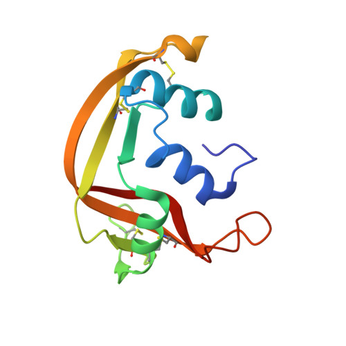
1GQV

Atomic Resolution (0.98A) Structure of Eosinophil-Derived Neurotoxin


Experimental Data Snapshot

  • Method: X-RAY DIFFRACTION
  • Resolution: 0.98 Å
  • R-Value Free: 0.144 
  • R-Value Observed: 0.116 

wwPDB Validation   3D Report Full Report


This is version 1.4 of the entry. See complete history


Literature

Atomic Resolution (0.98 A) Structure of Eosinophil-Derived Neurotoxin

Swaminathan, G.J.Holloway, D.E.Veluraja, K.Acharya, K.R.

(2002) Biochemistry 41: 3341

  • DOI: https://doi.org/10.1021/bi015911f
  • Primary Citation of Related Structures:  
    1GQV

  • PubMed Abstract: 

    Human eosinophil-derived neurotoxin (EDN) is a small, basic protein that belongs to the ribonuclease A superfamily. EDN displays antiviral activity and causes the neurotoxic Gordon phenomenon when injected into rabbits. Although EDN and ribonuclease A have appreciable structural similarity and a conserved catalytic triad, their peripheral substrate-binding sites are not conserved. The crystal structure of recombinant EDN (rEDN) has been determined at 0.98 A resolution from data collected at a low temperature (100 K). We have refined the crystallographic model of the structure using anisotropic displacement parameters to a conventional R-factor of 0.116. This represents the highest resolution structure of rEDN determined to date and is only the second ribonuclease structure to be determined at a resolution greater than 1.0 A. The structure provides a detailed picture of the conformational freedom at the various subsites of rEDN, and the water structure accounts for more than 50% of the total solvent content of the unit cell. This information will be crucial for the design of tight-binding inhibitors to restrain the ribonucleolytic activity of rEDN.


  • Organizational Affiliation

    Department of Biology and Biochemistry, University of Bath, Claverton Down, Bath BA2 7AY, U.K.


Macromolecules
Find similar proteins by:  (by identity cutoff)  |  3D Structure
Entity ID: 1
MoleculeChains Sequence LengthOrganismDetailsImage
EOSINOPHIL-DERIVED NEUROTOXIN135Homo sapiensMutation(s): 0 
EC: 3.1.27.5
UniProt & NIH Common Fund Data Resources
Find proteins for P10153 (Homo sapiens)
Explore P10153 
Go to UniProtKB:  P10153
PHAROS:  P10153
GTEx:  ENSG00000169385 
Entity Groups  
Sequence Clusters30% Identity50% Identity70% Identity90% Identity95% Identity100% Identity
UniProt GroupP10153
Sequence Annotations
Expand
  • Reference Sequence
Small Molecules
Ligands 1 Unique
IDChains Name / Formula / InChI Key2D Diagram3D Interactions
ACT
Query on ACT

Download Ideal Coordinates CCD File 
B [auth A]ACETATE ION
C2 H3 O2
QTBSBXVTEAMEQO-UHFFFAOYSA-M
Experimental Data & Validation

Experimental Data

  • Method: X-RAY DIFFRACTION
  • Resolution: 0.98 Å
  • R-Value Free: 0.144 
  • R-Value Observed: 0.116 
  • Space Group: P 21 21 21
Unit Cell:
Length ( Å )Angle ( ˚ )
a = 53.024α = 90
b = 57.477β = 90
c = 42.398γ = 90
Software Package:
Software NamePurpose
SHELXL-97refinement
DENZOdata reduction
SCALEPACKdata scaling
AMoREphasing
CNSphasing
SHELXphasing

Structure Validation

View Full Validation Report


Entry History 

Deposition Data

Revision History  (Full details and data files)

  • Version 1.0: 2002-03-08
    Type: Initial release
  • Version 1.1: 2011-05-08
    Changes: Version format compliance
  • Version 1.2: 2011-07-13
    Changes: Version format compliance
  • Version 1.3: 2019-05-22
    Changes: Data collection, Other, Refinement description
  • Version 1.4: 2023-12-13
    Changes: Data collection, Database references, Derived calculations, Refinement description