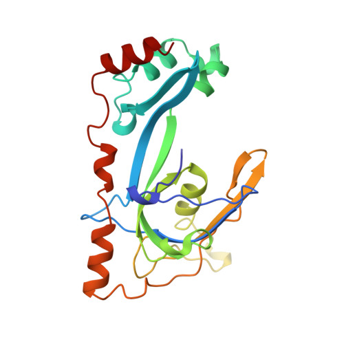
Crystal structure of a replication fork single-stranded DNA binding protein (T4 gp32) complexed to DNA.
Shamoo, Y., Friedman, A.M., Parsons, M.R., Konigsberg, W.H., Steitz, T.A.(1995) Nature 376: 362-366
- PubMed: 7630406
- DOI: https://doi.org/10.1038/376362a0
- Primary Citation of Related Structures:
1GPC - PubMed Abstract:
The single-stranded DNA (ssDNA) binding protein gp32 from bacteriophage T4 is essential for T4 DNA replication, recombination and repair. In vivo gp32 binds ssDNA as the replication fork advances and stimulates replisome processivity and accuracy by a factor of several hundred. Gp32 binding affects nearly every major aspect of DNA metabolism. Among its important functions are: (1) configuring ssDNA templates for efficient use by the replisome including DNA polymerase; (2) melting out adventitious secondary structures; (3) protecting exposed ssDNA from nucleases; and (4) facilitating homologous recombination by binding ssDNA during strand displacement. We have determined the crystal structure of the gp32 DNA binding domain complexed to ssDNA at 2.2 A resolution. The ssDNA binding cleft comprises regions from three structural subdomains and includes a positively charged surface that runs parallel to a series of hydrophobic pockets formed by clusters of aromatic side chains. Although only weak electron density is seen for the ssDNA, it indicates that the phosphate backbone contacts an electropositive cleft of the protein, placing the bases in contact with the hydrophobic pockets. The DNA mobility implied by the weak electron density may reflect the role of gp32 as a sequence-independent ssDNA chaperone allowing the largely unstructured ssDNA to slide freely through the cleft.
Organizational Affiliation:
Department of Molecular Biophysics and Biochemistry, Yale University, New Haven, Connecticut 06520-8114, USA.


