1GLH

CATION BINDING TO A BACILLUS (1,3-1,4)-BETA-GLUCANASE. GEOMETRY, AFFINITY AND EFFECT ON PROTEIN STABILITY


Experimental Data Snapshot

  • Method: X-RAY DIFFRACTION
  • Resolution: 2.00 Å
  • R-Value Free: 0.225 
  • R-Value Observed: 0.198 

wwPDB Validation   3D Report Full Report


This is version 1.2 of the entry. See complete history


Literature

Cation binding to a Bacillus (1,3-1,4)-beta-glucanase. Geometry, affinity and effect on protein stability

Keitel, T.Meldgaard, M.Heinemann, U.

(1994) Eur J Biochem 222: 203-214

  • DOI: https://doi.org/10.1111/j.1432-1033.1994.tb18858.x
  • Primary Citation of Related Structures:  
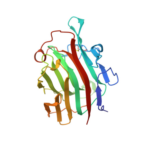
    1GLH

  • PubMed Abstract: 

    The hybrid Bacillus (1,3-1,4)-beta-glucanase H(A16-M), consisting of 16 N-terminal amino acids derived from the mature form of the B. amyloliquefaciens enzyme and of 198 C-proximal amino acids from the B. macerans enzyme, binds a calcium ion at a site at its molecular surface remote from the active center [T. Keitel, O. Simon, R. Borriss & U. Heinemann (1993) Proc. Natl Acad. Sci. USA 90, 5287-5291]. X-ray diffraction analysis at 0.22-nm resolution of crystals grown in the absence of calcium and in the presence of EDTA shows this site to be occupied by a sodium ion. Whereas the calcium ion has six oxygen atoms in its coordination sphere, two of which are from water molecules, sodium is fivefold coordinated with a fifth ligand belonging to a symmetry-related protein molecule in the crystal lattice. The affinity of H(A16-M) for calcium over sodium has been determined calorimetrically. Calcium binding stabilizes the native three-dimensional structure of the protein as shown by guanidinium chloride unfolding and thermal inactivation experiments. The enhanced enzymic activity of Bacillus beta-glucanases at elevated temperatures in the presence of calcium ions is attributed to a general stabilizing effect by the cation.


  • Organizational Affiliation

    Institut für Kristallographie, Freie Universität Berlin, Germany.


Macromolecules
Find similar proteins by:  (by identity cutoff)  |  3D Structure
Entity ID: 1
MoleculeChains Sequence LengthOrganismDetailsImage
1,3-1,4-BETA-GLUCANASE214synthetic constructMutation(s): 0 
EC: 3.2.1.73
UniProt
Find proteins for P23904 (Paenibacillus macerans)
Explore P23904 
Go to UniProtKB:  P23904
Entity Groups  
Sequence Clusters30% Identity50% Identity70% Identity90% Identity95% Identity100% Identity
UniProt GroupP23904
Sequence Annotations
Expand
  • Reference Sequence
Small Molecules
Ligands 1 Unique
IDChains Name / Formula / InChI Key2D Diagram3D Interactions
NA
Query on NA

Download Ideal Coordinates CCD File 
B [auth A]SODIUM ION
Na
FKNQFGJONOIPTF-UHFFFAOYSA-N
Experimental Data & Validation

Experimental Data

  • Method: X-RAY DIFFRACTION
  • Resolution: 2.00 Å
  • R-Value Free: 0.225 
  • R-Value Observed: 0.198 
  • Space Group: P 21 21 21
Unit Cell:
Length ( Å )Angle ( ˚ )
a = 70.22α = 90
b = 72.56β = 90
c = 49.97γ = 90
Software Package:
Software NamePurpose
PROFFTrefinement

Structure Validation

View Full Validation Report


Entry History 

Deposition Data

Revision History  (Full details and data files)

  • Version 1.0: 1995-02-07
    Type: Initial release
  • Version 1.1: 2008-03-24
    Changes: Version format compliance
  • Version 1.2: 2011-07-13
    Changes: Version format compliance