1GCM

GCN4 LEUCINE ZIPPER CORE MUTANT P-LI


Experimental Data Snapshot

  • Method: X-RAY DIFFRACTION
  • Resolution: 1.80 Å
  • R-Value Observed: 0.175 

wwPDB Validation   3D Report Full Report


This is version 1.3 of the entry. See complete history


Literature

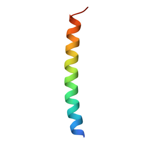
Crystal structure of an isoleucine-zipper trimer.

Harbury, P.B.Kim, P.S.Alber, T.

(1994) Nature 371: 80-83

  • DOI: https://doi.org/10.1038/371080a0
  • Primary Citation of Related Structures:  
    1GCM

  • PubMed Abstract: 

    Subunit oligomerization in many proteins is mediated by short coiled-coil motifs. These motifs share a characteristic seven-amino-acid repeat containing hydrophobic residues at the first (a) and fourth (d) positions. Despite this common pattern, different sequences form two-, three- and four-stranded helical ropes. We have investigated the basis for oligomer choice by characterizing variants of the GCN4 leucine-zipper dimerization domain that adopt trimeric or tetrameric structures in response to mutations at the a and d positions. We now report the high-resolution X-ray crystal structure of an isoleucine-containing mutant that folds into a parallel three-stranded, alpha-helical coiled coil. In contrast to the dimer and tetramer structures, the interior packing of the trimer can accommodate beta-branched residues in the most preferred rotamer at both hydrophobic positions. Compatibility of the shape of the core amino acids with the distinct packing spaces in the two-, three- and four-stranded conformations appears to determine the oligomerization state of the GCN4 leucine-zipper variants.


  • Organizational Affiliation

    Department of Biological Chemistry and Molecular Pharmacology, Harvard Medical School, Boston, Massachusetts 02115.


Macromolecules
Find similar proteins by:  (by identity cutoff)  |  3D Structure
Entity ID: 1
MoleculeChains Sequence LengthOrganismDetailsImage
GCN4P-II
A, B, C
34Saccharomyces cerevisiaeMutation(s): 8 
UniProt
Find proteins for P03069 (Saccharomyces cerevisiae (strain ATCC 204508 / S288c))
Explore P03069 
Go to UniProtKB:  P03069
Entity Groups  
Sequence Clusters30% Identity50% Identity70% Identity90% Identity95% Identity100% Identity
UniProt GroupP03069
Sequence Annotations
Expand
  • Reference Sequence
Experimental Data & Validation

Experimental Data

  • Method: X-RAY DIFFRACTION
  • Resolution: 1.80 Å
  • R-Value Observed: 0.175 
  • Space Group: P 1 21 1
Unit Cell:
Length ( Å )Angle ( ˚ )
a = 35.32α = 90
b = 39.67β = 98.38
c = 36.97γ = 90
Software Package:
Software NamePurpose
X-PLORmodel building
TNTrefinement
X-PLORrefinement
R-AXISdata reduction
X-PLORphasing

Structure Validation

View Full Validation Report


Entry History 

Deposition Data

Revision History  (Full details and data files)

  • Version 1.0: 1996-01-29
    Type: Initial release
  • Version 1.1: 2008-03-24
    Changes: Version format compliance
  • Version 1.2: 2011-07-13
    Changes: Version format compliance
  • Version 1.3: 2021-11-03
    Changes: Database references, Derived calculations, Other