1GCL

GCN4 LEUCINE ZIPPER CORE MUTANT P-LI


Experimental Data Snapshot

  • Method: X-RAY DIFFRACTION
  • Resolution: 2.10 Å
  • R-Value Observed: 0.152 

wwPDB Validation   3D Report Full Report


This is version 1.3 of the entry. See complete history


Literature

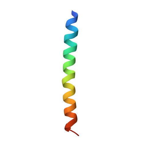
A switch between two-, three-, and four-stranded coiled coils in GCN4 leucine zipper mutants.

Harbury, P.B.Zhang, T.Kim, P.S.Alber, T.

(1993) Science 262: 1401-1407

  • DOI: https://doi.org/10.1126/science.8248779
  • Primary Citation of Related Structures:  
    1GCL

  • PubMed Abstract: 

    Coiled-coil sequences in proteins consist of heptad repeats containing two characteristic hydrophobic positions. The role of these buried hydrophobic residues in determining the structures of coiled coils was investigated by studying mutants of the GCN4 leucine zipper. When sets of buried residues were altered, two-, three-, and four-helix structures were formed. The x-ray crystal structure of the tetramer revealed a parallel, four-stranded coiled coil. In the tetramer conformation, the local packing geometry of the two hydrophobic positions in the heptad repeat is reversed relative to that in the dimer. These studies demonstrate that conserved, buried residues in the GCN4 leucine zipper direct dimer formation. In contrast to proposals that the pattern of hydrophobic and polar amino acids in a protein sequence is sufficient to determine three-dimensional structure, the shapes of buried side chains in coiled coils are essential determinants of the global fold.


  • Organizational Affiliation

    Department of Biological Chemistry and Molecular Pharmacology, Harvard Medical School, Boston, MA 02115.


Macromolecules
Find similar proteins by:  (by identity cutoff)  |  3D Structure
Entity ID: 1
MoleculeChains Sequence LengthOrganismDetailsImage
GCN4
A, B, C, D
34Saccharomyces cerevisiaeMutation(s): 8 
UniProt
Find proteins for P03069 (Saccharomyces cerevisiae (strain ATCC 204508 / S288c))
Explore P03069 
Go to UniProtKB:  P03069
Entity Groups  
Sequence Clusters30% Identity50% Identity70% Identity90% Identity95% Identity100% Identity
UniProt GroupP03069
Sequence Annotations
Expand
  • Reference Sequence
Experimental Data & Validation

Experimental Data

  • Method: X-RAY DIFFRACTION
  • Resolution: 2.10 Å
  • R-Value Observed: 0.152 
  • Space Group: P 21 21 21
Unit Cell:
Length ( Å )Angle ( ˚ )
a = 47.432α = 90
b = 48.634β = 90
c = 51.765γ = 90
Software Package:
Software NamePurpose
X-PLORmodel building
TNTrefinement
X-PLORrefinement
R-AXISdata reduction
X-PLORphasing

Structure Validation

View Full Validation Report


Entry History 

Deposition Data

Revision History  (Full details and data files)

  • Version 1.0: 1995-06-03
    Type: Initial release
  • Version 1.1: 2008-03-24
    Changes: Version format compliance
  • Version 1.2: 2011-07-13
    Changes: Version format compliance
  • Version 1.3: 2013-04-24
    Changes: Other