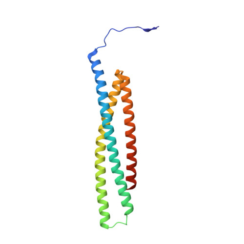
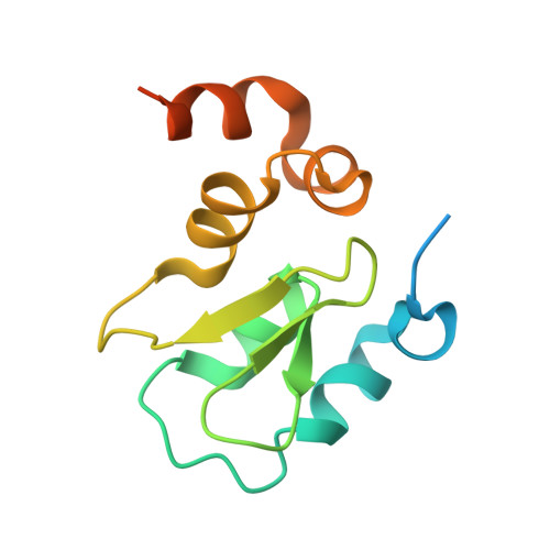
1G73

CRYSTAL STRUCTURE OF SMAC BOUND TO XIAP-BIR3 DOMAIN


Experimental Data Snapshot

  • Method: X-RAY DIFFRACTION
  • Resolution: 2.00 Å
  • R-Value Free: 0.268 
  • R-Value Work: 0.227 
  • R-Value Observed: 0.227 

wwPDB Validation   3D Report Full Report


This is version 1.4 of the entry. See complete history


Literature

Structural basis of IAP recognition by Smac/DIABLO.

Wu, G.Chai, J.Suber, T.L.Wu, J.W.Du, C.Wang, X.Shi, Y.

(2000) Nature 408: 1008-1012

  • DOI: https://doi.org/10.1038/35050012
  • Primary Citation of Related Structures:  
    1G73

  • PubMed Abstract: 

    Apoptosis is an essential process in the development and homeostasis of all metazoans. The inhibitor-of-apoptosis (IAP) proteins suppress cell death by inhibiting the activity of caspases; this inhibition is performed by the zinc-binding BIR domains of the IAP proteins. The mitochondrial protein Smac/DIABLO promotes apoptosis by eliminating the inhibitory effect of IAPs through physical interactions. Amino-terminal sequences in Smac/DIABLO are required for this function, as mutation of the very first amino acid leads to loss of interaction with IAPs and concomitant loss of Smac/DIABLO function. Here we report the high-resolution crystal structure of Smac/DIABLO complexed with the third BIR domain (BIR3) of XIAP. Our results show that the N-terminal four residues (Ala-Val-Pro-Ile) in Smac/DIABLO recognize a surface groove on BIR3, with the first residue Ala binding a hydrophobic pocket and making five hydrogen bonds to neighbouring residues on BIR3. These observations provide a structural explanation for the roles of the Smac N terminus as well as the conserved N-terminal sequences in the Drosophila proteins Hid/Grim/Reaper. In conjunction with other observations, our results reveal how Smac may relieve IAP inhibition of caspase-9 activity. In addition to explaining a number of biological observations, our structural analysis identifies potential targets for drug screening.


  • Organizational Affiliation

    Department of Molecular Biology, Princeton University, New Jersey 08544, USA.


Macromolecules
Find similar proteins by:  (by identity cutoff)  |  3D Structure
Entity ID: 1
MoleculeChains Sequence LengthOrganismDetailsImage
SECOND MITOCHONDRIA-DERIVED ACTIVATOR OF CASPASES
A, B
162Homo sapiensMutation(s): 1 
Gene Names: SMAC
UniProt & NIH Common Fund Data Resources
Find proteins for Q9NR28 (Homo sapiens)
Explore Q9NR28 
Go to UniProtKB:  Q9NR28
PHAROS:  Q9NR28
GTEx:  ENSG00000184047 
Entity Groups  
Sequence Clusters30% Identity50% Identity70% Identity90% Identity95% Identity100% Identity
UniProt GroupQ9NR28
Sequence Annotations
Expand
  • Reference Sequence
Find similar proteins by:  (by identity cutoff)  |  3D Structure
Entity ID: 2
MoleculeChains Sequence LengthOrganismDetailsImage
INHIBITORS OF APOPTOSIS-LIKE PROTEIN ILP
C, D
121Homo sapiensMutation(s): 0 
UniProt & NIH Common Fund Data Resources
Find proteins for P98170 (Homo sapiens)
Explore P98170 
Go to UniProtKB:  P98170
PHAROS:  P98170
GTEx:  ENSG00000101966 
Entity Groups  
Sequence Clusters30% Identity50% Identity70% Identity90% Identity95% Identity100% Identity
UniProt GroupP98170
Sequence Annotations
Expand
  • Reference Sequence
Small Molecules
Ligands 1 Unique
IDChains Name / Formula / InChI Key2D Diagram3D Interactions
ZN
Query on ZN

Download Ideal Coordinates CCD File 
E [auth B],
F [auth D]
ZINC ION
Zn
PTFCDOFLOPIGGS-UHFFFAOYSA-N
Experimental Data & Validation

Experimental Data

  • Method: X-RAY DIFFRACTION
  • Resolution: 2.00 Å
  • R-Value Free: 0.268 
  • R-Value Work: 0.227 
  • R-Value Observed: 0.227 
  • Space Group: P 1
Unit Cell:
Length ( Å )Angle ( ˚ )
a = 48.1α = 100
b = 52.9β = 104.1
c = 67.1γ = 94
Software Package:
Software NamePurpose
MLPHAREphasing
X-PLORrefinement
DENZOdata reduction
SCALEPACKdata scaling

Structure Validation

View Full Validation Report


Entry History 

Deposition Data

Revision History  (Full details and data files)

  • Version 1.0: 2001-01-10
    Type: Initial release
  • Version 1.1: 2008-04-27
    Changes: Version format compliance
  • Version 1.2: 2011-07-13
    Changes: Version format compliance
  • Version 1.3: 2021-10-27
    Changes: Database references, Derived calculations
  • Version 1.4: 2024-02-07
    Changes: Data collection