1FZR

CRYSTAL STRUCTURE OF BACTERIOPHAGE T7 ENDONUCLEASE I


Experimental Data Snapshot

  • Method: X-RAY DIFFRACTION
  • Resolution: 2.10 Å
  • R-Value Free: 0.235 
  • R-Value Work: 0.198 
  • R-Value Observed: 0.202 

wwPDB Validation   3D Report Full Report


This is version 1.4 of the entry. See complete history


Literature

Crystal structure of the Holliday junction resolving enzyme T7 endonuclease I.

Hadden, J.M.Convery, M.A.Declais, A.C.Lilley, D.M.Phillips, S.E.

(2001) Nat Struct Biol 8: 62-67

  • DOI: https://doi.org/10.1038/83067
  • Primary Citation of Related Structures:  
    1FZR

  • PubMed Abstract: 

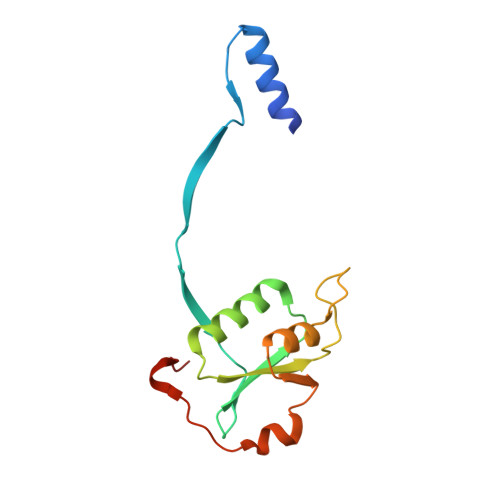
    We have solved the crystal structure of the Holliday junction resolving enzyme T7 endonuclease I at 2.1 A resolution using the multiwavelength anomalous dispersion (MAD) technique. Endonuclease I exhibits strong structural specificity for four-way DNA junctions. The structure shows that it forms a symmetric homodimer arranged in two well-separated domains. Each domain, however, is composed of elements from both subunits, and amino acid side chains from both protomers contribute to the active site. While no significant structural similarity could be detected with any other junction resolving enzyme, the active site is similar to that found in several restriction endonucleases. T7 endonuclease I therefore represents the first crystal structure of a junction resolving enzyme that is a member of the nuclease superfamily of enzymes.


  • Organizational Affiliation

    Astbury Centre for Structural Molecular Biology, School of Biochemistry and Molecular Biology, University of Leeds, Leeds LS2 9JT, UK.


Macromolecules
Find similar proteins by:  (by identity cutoff)  |  3D Structure
Entity ID: 1
MoleculeChains Sequence LengthOrganismDetailsImage
ENDONUCLEASE I
A, B, C, D
138Escherichia phage T7Mutation(s): 1 
EC: 3.1.21.2
UniProt
Find proteins for P00641 (Escherichia phage T7)
Explore P00641 
Go to UniProtKB:  P00641
Entity Groups  
Sequence Clusters30% Identity50% Identity70% Identity90% Identity95% Identity100% Identity
UniProt GroupP00641
Sequence Annotations
Expand
  • Reference Sequence
Experimental Data & Validation

Experimental Data

  • Method: X-RAY DIFFRACTION
  • Resolution: 2.10 Å
  • R-Value Free: 0.235 
  • R-Value Work: 0.198 
  • R-Value Observed: 0.202 
  • Space Group: P 21 21 2
Unit Cell:
Length ( Å )Angle ( ˚ )
a = 123.66α = 90
b = 135.19β = 90
c = 61.58γ = 90
Software Package:
Software NamePurpose
SOLVEphasing
CNSrefinement
MOSFLMdata reduction
CCP4data scaling

Structure Validation

View Full Validation Report


Entry History 

Deposition Data

Revision History  (Full details and data files)

  • Version 1.0: 2001-01-17
    Type: Initial release
  • Version 1.1: 2008-04-27
    Changes: Version format compliance
  • Version 1.2: 2011-07-13
    Changes: Version format compliance
  • Version 1.3: 2021-11-03
    Changes: Database references, Derived calculations
  • Version 1.4: 2024-02-07
    Changes: Data collection