1FX4

STRUCTURE ANALYSIS OF ADENYLATE CYCLASES FROM TRYPANOSOMA BRUCEI IN THEIR MONOMERIC STATE


Experimental Data Snapshot

  • Method: X-RAY DIFFRACTION
  • Resolution: 1.90 Å
  • R-Value Free: 0.232 
  • R-Value Work: 0.202 
  • R-Value Observed: 0.202 

wwPDB Validation   3D Report Full Report


This is version 1.4 of the entry. See complete history


Literature

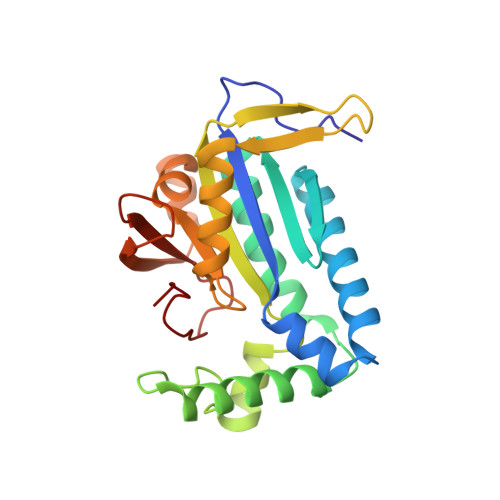
Structural analysis of adenylate cyclases from Trypanosoma brucei in their monomeric state.

Bieger, B.Essen, L.O.

(2001) EMBO J 20: 433-445

  • DOI: https://doi.org/10.1093/emboj/20.3.433
  • Primary Citation of Related Structures:  
    1FX2, 1FX4

  • PubMed Abstract: 

    Cyclic AMP is a major trigger of the differentiation process of Trypanosoma brucei, a bloodstream parasite causing sleeping sickness. Its generation in trypanosomes is accomplished by a unique battery of membrane-bound adenylate cyclases (ACs). We have determined the high-resolution X-ray structures of the catalytic domains of two trypanosomal ACs (tACs), GRESAG4.1 and GRESAG4.3. The tAC domains are structurally highly related to the AC domains of higher eukaryotes, but also comprise a highly conserved structural element near the active site, the Delta-subdomain. A cavity below the Delta-subdomain might correspond to an allosteric regulator site as indicated by the stereospecific binding of a single (2S,3S)-1,4- dimercapto-2,3-butanediol molecule. In three different crystal forms, the tAC domains are exclusively observed in a monomeric, catalytically inactive state. Biochemical analysis and the mutagenesis profile of GRESAG4.1 confirmed a common catalytic mechanism of tACs that involves transient dimerization of the AC domain. A low dimerization tendency might play a regulatory role in T. brucei if the activation of tACs is similarly driven by ligand-induced dimerization as in membrane-bound guanylate cyclases.


  • Organizational Affiliation

    Max-Planck-Institute for Biochemistry, Department of Membrane Biochemistry, Am Klopferspitz 18a, D-82152 Martinsried bei München, Germany.


Macromolecules
Find similar proteins by:  (by identity cutoff)  |  3D Structure
Entity ID: 1
MoleculeChains Sequence LengthOrganismDetailsImage
RECEPTOR-TYPE ADENYLATE CYCLASE GRESAG 4.3231Trypanosoma bruceiMutation(s): 0 
EC: 4.6.1.1
UniProt
Find proteins for Q99280 (Trypanosoma brucei brucei)
Explore Q99280 
Go to UniProtKB:  Q99280
Entity Groups  
Sequence Clusters30% Identity50% Identity70% Identity90% Identity95% Identity100% Identity
UniProt GroupQ99280
Sequence Annotations
Expand
  • Reference Sequence
Small Molecules
Ligands 1 Unique
IDChains Name / Formula / InChI Key2D Diagram3D Interactions
MG
Query on MG

Download Ideal Coordinates CCD File 
B [auth A]MAGNESIUM ION
Mg
JLVVSXFLKOJNIY-UHFFFAOYSA-N
Experimental Data & Validation

Experimental Data

  • Method: X-RAY DIFFRACTION
  • Resolution: 1.90 Å
  • R-Value Free: 0.232 
  • R-Value Work: 0.202 
  • R-Value Observed: 0.202 
  • Space Group: I 41
Unit Cell:
Length ( Å )Angle ( ˚ )
a = 90α = 90
b = 90β = 90
c = 66.9γ = 90
Software Package:
Software NamePurpose
DENZOdata reduction
SCALEPACKdata scaling
AMoREphasing
CNSrefinement

Structure Validation

View Full Validation Report


Entry History 

Deposition Data

Revision History  (Full details and data files)

  • Version 1.0: 2001-02-28
    Type: Initial release
  • Version 1.1: 2008-04-27
    Changes: Version format compliance
  • Version 1.2: 2011-07-13
    Changes: Version format compliance
  • Version 1.3: 2018-01-31
    Changes: Advisory, Experimental preparation
  • Version 1.4: 2024-02-07
    Changes: Advisory, Data collection, Database references, Derived calculations