1FW4

CRYSTAL STRUCTURE OF E. COLI FRAGMENT TR2C FROM CALMODULIN TO 1.7 A RESOLUTION


Experimental Data Snapshot

  • Method: X-RAY DIFFRACTION
  • Resolution: 1.70 Å
  • R-Value Free: 0.220 
  • R-Value Work: 0.218 

wwPDB Validation   3D Report Full Report


This is version 1.4 of the entry. See complete history


Literature

Structure of Escherichia coli fragment TR2C from calmodulin to 1.7 A resolution.

Olsson, L.L.Sjolin, L.

(2001) Acta Crystallogr D Biol Crystallogr 57: 664-669

  • DOI: https://doi.org/10.1107/s090744490100347x
  • Primary Citation of Related Structures:  
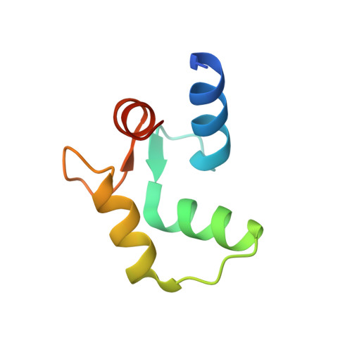
    1FW4

  • PubMed Abstract: 

    Fragment TR2C is the C-terminal part of the calcium-binding protein calmodulin, including residues 78-148. The crystal structure of TR2C was solved by molecular replacement and refined to a conventional R value of 21.8% (R(free) = 22.0%), using all data in the resolution range 20.0-1.7 A. This study shows that the secondary structure of TR2C, a pair of EF-hand motifs with two calcium-binding sites, is similar to the corresponding motifs in intact calmodulin. However, it also indicates that the N-terminus of helix E is closer to the C-terminus of helix H in TR2C than in the intact protein and that the loop connecting the EF-hands shows different conformations in the two structures. The crystal structure of TR2C was further found to be similar to the set of NMR structures of this fragment, although some pronounced differences exist.


  • Organizational Affiliation

    Department of Inorganic Chemistry and the Center for Structural Biology, Göteborg University, SE-412 96 Göteborg, Sweden.


Macromolecules
Find similar proteins by:  (by identity cutoff)  |  3D Structure
Entity ID: 1
MoleculeChains Sequence LengthOrganismDetailsImage
CALMODULIN71Bos taurusMutation(s): 0 
UniProt
Find proteins for P62157 (Bos taurus)
Explore P62157 
Go to UniProtKB:  P62157
Entity Groups  
Sequence Clusters30% Identity50% Identity70% Identity90% Identity95% Identity100% Identity
UniProt GroupP62157
Sequence Annotations
Expand
  • Reference Sequence
Experimental Data & Validation

Experimental Data

  • Method: X-RAY DIFFRACTION
  • Resolution: 1.70 Å
  • R-Value Free: 0.220 
  • R-Value Work: 0.218 
  • Space Group: I 41
Unit Cell:
Length ( Å )Angle ( ˚ )
a = 37.802α = 90
b = 37.802β = 90
c = 99.779γ = 90
Software Package:
Software NamePurpose
AMoREphasing
CNSrefinement
DENZOdata reduction
SCALEPACKdata scaling

Structure Validation

View Full Validation Report


Entry History 

Deposition Data

Revision History  (Full details and data files)

  • Version 1.0: 2001-05-02
    Type: Initial release
  • Version 1.1: 2008-04-27
    Changes: Version format compliance
  • Version 1.2: 2011-07-13
    Changes: Version format compliance
  • Version 1.3: 2018-03-07
    Changes: Data collection
  • Version 1.4: 2023-08-09
    Changes: Data collection, Database references, Derived calculations, Refinement description