1FOB

CRYSTAL STRUCTURE OF BETA-1,4-GALACTANASE FROM ASPERGILLUS ACULEATUS AT 100K


Experimental Data Snapshot

  • Method: X-RAY DIFFRACTION
  • Resolution: 1.80 Å
  • R-Value Free: 0.251 
  • R-Value Work: 0.211 
  • R-Value Observed: 0.211 

wwPDB Validation   3D Report Full Report


This is version 1.3 of the entry. See complete history


Literature

Aspergillus aculeatus beta-1,4-Galactanase: Substrate Recognition and Relations to Other Glycoside Hydrolases in Clan GH-A

Ryttersgaard, C.Leggio, L.L.Coutinho, P.M.Henrissat, B.Larsen, S.

(2002) Biochemistry 41: 15135-15143

  • DOI: https://doi.org/10.1021/bi026238c
  • Primary Citation of Related Structures:  
    1FHL, 1FOB

  • PubMed Abstract: 

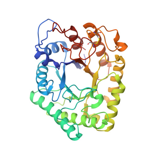
    The three-dimensional structure of Aspergillus aculeatus beta-1,4-galactanase (AAGAL), an enzyme involved in pectin degradation, has been determined by multiple isomorphous replacement to 2.3 and 1.8 A resolution at 293 and 100 K, respectively. It represents the first known structure for a polysaccharidase with this specificity and for a member of glycoside hydrolase family 53 (GH-53). The enzyme folds into a (beta/alpha)(8) barrel with the active site cleft located at the C-terminal side of the barrel consistent with the classification of GH-53 in clan GH-A, a superfamily of enzymes with common fold and catalytic machinery but diverse specificities. Putative substrate-enzyme interactions were elucidated by modeling of beta-1,4-linked galactobioses into the possible substrate binding subsites. The structure and modeling studies identified five potential subsites for the binding of galactans, of which one is a pocket suited for accommodating the arabinan side chain in arabinogalactan, one of the natural substrates. A comparison with the substrate binding grooves of other Clan GH-A enzymes suggests that shape complementarity is crucial in determining the specificity of polysaccharidases.


  • Organizational Affiliation

    Centre for Crystallographic Studies, Department of Chemistry, University of Copenhagen, Universitetsparken 5, DK-2100 Copenhagen, Denmark.


Macromolecules
Find similar proteins by:  (by identity cutoff)  |  3D Structure
Entity ID: 1
MoleculeChains Sequence LengthOrganismDetailsImage
BETA-1,4-GALACTANASE334Aspergillus aculeatusMutation(s): 0 
EC: 3.2.1.89
UniProt
Find proteins for P48842 (Aspergillus aculeatus)
Explore P48842 
Go to UniProtKB:  P48842
Entity Groups  
Sequence Clusters30% Identity50% Identity70% Identity90% Identity95% Identity100% Identity
UniProt GroupP48842
Sequence Annotations
Expand
  • Reference Sequence
Small Molecules
Ligands 1 Unique
IDChains Name / Formula / InChI Key2D Diagram3D Interactions
CA
Query on CA

Download Ideal Coordinates CCD File 
B [auth A]CALCIUM ION
Ca
BHPQYMZQTOCNFJ-UHFFFAOYSA-N
Experimental Data & Validation

Experimental Data

  • Method: X-RAY DIFFRACTION
  • Resolution: 1.80 Å
  • R-Value Free: 0.251 
  • R-Value Work: 0.211 
  • R-Value Observed: 0.211 
  • Space Group: I 2 2 2
Unit Cell:
Length ( Å )Angle ( ˚ )
a = 59.7α = 90
b = 87.98β = 90
c = 128.2γ = 90
Software Package:
Software NamePurpose
MAR345data collection
SCALEPACKdata scaling
CNSrefinement
CNSphasing

Structure Validation

View Full Validation Report


Entry History 

Deposition Data

Revision History  (Full details and data files)

  • Version 1.0: 2003-06-03
    Type: Initial release
  • Version 1.1: 2008-04-27
    Changes: Version format compliance
  • Version 1.2: 2011-07-13
    Changes: Version format compliance
  • Version 1.3: 2017-10-04
    Changes: Data collection, Refinement description