1FIB

RECOMBINANT HUMAN GAMMA-FIBRINOGEN CARBOXYL TERMINAL FRAGMENT (RESIDUES 143-411) BOUND TO CALCIUM AT PH 6.0


Experimental Data Snapshot

  • Method: X-RAY DIFFRACTION
  • Resolution: 2.10 Å
  • R-Value Free: 0.227 
  • R-Value Work: 0.159 
  • R-Value Observed: 0.159 

wwPDB Validation   3D Report Full Report


This is version 1.2 of the entry. See complete history


Literature

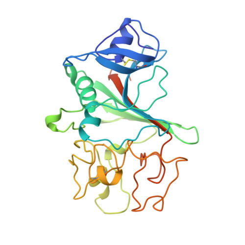
Crystal structure of a 30 kDa C-terminal fragment from the gamma chain of human fibrinogen.

Yee, V.C.Pratt, K.P.Cote, H.C.Trong, I.L.Chung, D.W.Davie, E.W.Stenkamp, R.E.Teller, D.C.

(1997) Structure 5: 125-138

  • DOI: https://doi.org/10.1016/s0969-2126(97)00171-8
  • Primary Citation of Related Structures:  
    1FIB, 1FIC, 1FID

  • PubMed Abstract: 

    Blood coagulation occurs by a cascade of zymogen activation resulting from minor proteolysis. The final stage of coagulation involves thrombin generation and limited proteolysis of fibrinogen to give spontaneously polymerizing fibrin. The resulting fibrin network is covalently crosslinked by factor XIIIa to yield a stable blood clot. Fibrinogen is a 340 kDa glycoprotein composed of six polypeptide chains, (alphabetagamma)2, held together by 29 disulfide bonds. The globular C terminus of the gamma chain contains a fibrin-polymerization surface, the principal factor XIIIa crosslinking site, the platelet receptor recognition site, and a calcium-binding site. Structural information on this domain should thus prove helpful in understanding clot formation.


  • Organizational Affiliation

    Department of Biochemistry, Biomolecular Structure Center, University of Washington, Seattle, WA 98195, USA.


Macromolecules
Find similar proteins by:  (by identity cutoff)  |  3D Structure
Entity ID: 1
MoleculeChains Sequence LengthOrganismDetailsImage
GAMMA-FIBRINOGEN CARBOXYL TERMINAL FRAGMENT269Homo sapiensMutation(s): 0 
Gene Names: HUMAN FIBRINOGEN GAMMA CHAIN C
UniProt & NIH Common Fund Data Resources
Find proteins for P02679 (Homo sapiens)
Explore P02679 
Go to UniProtKB:  P02679
PHAROS:  P02679
GTEx:  ENSG00000171557 
Entity Groups  
Sequence Clusters30% Identity50% Identity70% Identity90% Identity95% Identity100% Identity
UniProt GroupP02679
Sequence Annotations
Expand
  • Reference Sequence
Small Molecules
Ligands 1 Unique
IDChains Name / Formula / InChI Key2D Diagram3D Interactions
CA
Query on CA

Download Ideal Coordinates CCD File 
B [auth A]CALCIUM ION
Ca
BHPQYMZQTOCNFJ-UHFFFAOYSA-N
Experimental Data & Validation

Experimental Data

  • Method: X-RAY DIFFRACTION
  • Resolution: 2.10 Å
  • R-Value Free: 0.227 
  • R-Value Work: 0.159 
  • R-Value Observed: 0.159 
  • Space Group: P 1 21 1
Unit Cell:
Length ( Å )Angle ( ˚ )
a = 37.53α = 90
b = 68.42β = 105.36
c = 47.67γ = 90
Software Package:
Software NamePurpose
XENGENdata collection
X-PLORmodel building
X-PLORrefinement
XENGENdata reduction
X-PLORphasing

Structure Validation

View Full Validation Report


Entry History 

Deposition Data

Revision History  (Full details and data files)

  • Version 1.0: 1997-04-01
    Type: Initial release
  • Version 1.1: 2008-03-24
    Changes: Version format compliance
  • Version 1.2: 2011-07-13
    Changes: Version format compliance