1FI8

RAT GRANZYME B [N66Q] COMPLEXED TO ECOTIN [81-84 IEPD]


Experimental Data Snapshot

  • Method: X-RAY DIFFRACTION
  • Resolution: 2.20 Å
  • R-Value Free: 0.252 
  • R-Value Work: 0.218 
  • R-Value Observed: 0.222 

wwPDB Validation   3D Report Full Report


This is version 1.3 of the entry. See complete history


Literature

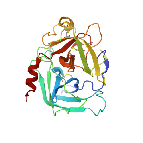
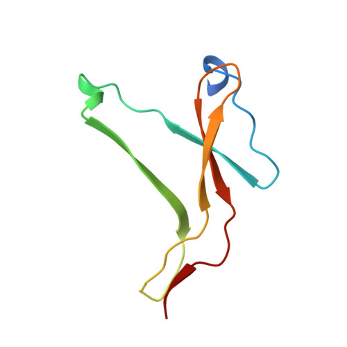
The structure of the pro-apoptotic protease granzyme B reveals the molecular determinants of its specificity

Waugh, S.M.Harris, J.L.Fletterick, R.Craik, C.S.

(2000) Nat Struct Biol 7: 762-765

  • DOI: https://doi.org/10.1038/78992
  • Primary Citation of Related Structures:  
    1FI8

  • PubMed Abstract: 

    Granzyme B is a serine protease of the chymotrypsin fold that mediates cell death by cytotoxic lymphocytes. It is a processing enzyme, requiring extended peptide substrates containing an Asp residue. The determinants that allow for this substrate specificity are revealed in the three-dimensional structure of granzyme B in complex with a macromolecular inhibitor. The primary specificity for Asp occurs through a side-on interaction with Arg 226, a buried Arg side chain of granzyme B. An additional nine amino acids make contact with the substrate and define the granzyme B extended substrate specificity profile. The substrate determinants found in this structure are shared by other members of this protein class and help to reveal the properties that define substrate specificity.


  • Organizational Affiliation

    The Graduate Group in Biophysics, University of California, San Francisco, California 94143-0446, USA. waugh@mutant.ucsf.edu


Macromolecules
Find similar proteins by:  (by identity cutoff)  |  3D Structure
Entity ID: 1
MoleculeChains Sequence LengthOrganismDetailsImage
NATURAL KILLER CELL PROTEASE 1A,
D [auth B]
228Rattus norvegicusMutation(s): 1 
EC: 3.4.21
UniProt
Find proteins for P18291 (Rattus norvegicus)
Explore P18291 
Go to UniProtKB:  P18291
Entity Groups  
Sequence Clusters30% Identity50% Identity70% Identity90% Identity95% Identity100% Identity
UniProt GroupP18291
Sequence Annotations
Expand
  • Reference Sequence
Find similar proteins by:  (by identity cutoff)  |  3D Structure
Entity ID: 2
MoleculeChains Sequence LengthOrganismDetailsImage
ECOTINB [auth C],
E
84Escherichia coliMutation(s): 4 
UniProt
Find proteins for P23827 (Escherichia coli (strain K12))
Explore P23827 
Go to UniProtKB:  P23827
Entity Groups  
Sequence Clusters30% Identity50% Identity70% Identity90% Identity95% Identity100% Identity
UniProt GroupP23827
Sequence Annotations
Expand
  • Reference Sequence
Find similar proteins by:  (by identity cutoff)  |  3D Structure
Entity ID: 3
MoleculeChains Sequence LengthOrganismDetailsImage
ECOTINC [auth D],
F
58Escherichia coliMutation(s): 0 
UniProt
Find proteins for P23827 (Escherichia coli (strain K12))
Explore P23827 
Go to UniProtKB:  P23827
Entity Groups  
Sequence Clusters30% Identity50% Identity70% Identity90% Identity95% Identity100% Identity
UniProt GroupP23827
Sequence Annotations
Expand
  • Reference Sequence
Experimental Data & Validation

Experimental Data

  • Method: X-RAY DIFFRACTION
  • Resolution: 2.20 Å
  • R-Value Free: 0.252 
  • R-Value Work: 0.218 
  • R-Value Observed: 0.222 
  • Space Group: P 1 21 1
Unit Cell:
Length ( Å )Angle ( ˚ )
a = 56.64α = 90
b = 154.6β = 119.76
c = 57.24γ = 90
Software Package:
Software NamePurpose
MOSFLMdata reduction
SCALAdata scaling
EPMRphasing
CNSrefinement
CCP4data scaling

Structure Validation

View Full Validation Report


Entry History 

Deposition Data

Revision History  (Full details and data files)

  • Version 1.0: 2000-09-13
    Type: Initial release
  • Version 1.1: 2008-04-27
    Changes: Version format compliance
  • Version 1.2: 2011-07-13
    Changes: Version format compliance
  • Version 1.3: 2021-11-03
    Changes: Database references