1FE6

CRYSTAL STRUCTURE OF A NATURALLY OCCURING PARALLEL RIGHT-HANDED COILED-COIL TETRAMER


Experimental Data Snapshot

  • Method: X-RAY DIFFRACTION
  • Resolution: 1.80 Å
  • R-Value Free: 0.216 
  • R-Value Work: 0.198 
  • R-Value Observed: 0.198 

wwPDB Validation   3D Report Full Report


This is version 1.3 of the entry. See complete history


Literature

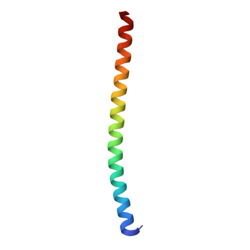
Crystal structure of a naturally occurring parallel right-handed coiled coil tetramer.

Stetefeld, J.Jenny, M.Schulthess, T.Landwehr, R.Engel, J.Kammerer, R.A.

(2000) Nat Struct Biol 7: 772-776

  • DOI: https://doi.org/10.1038/79006
  • Primary Citation of Related Structures:  
    1FE6

  • PubMed Abstract: 

    The crystal structure of a polypeptide chain fragment from the surface layer protein tetrabrachion from Staphylothermus marinus has been determined at 1.8 A resolution. As proposed on the basis of the presence of 11-residue repeats, the polypeptide chain fragment forms a parallel right-handed coiled coil structure. Complementary hydrophobic interactions and complex networks of surface salt bridges result in an extremely thermostable tetrameric structure with remarkable properties. In marked contrast to left-handed coiled coil tetramers, the right-handed coiled coil reveals large hydrophobic cavities that are filled with water molecules. As a consequence, the packing of the hydrophobic core differs markedly from that of a right-handed parallel coiled coil tetramer that was designed on the basis of left-handed coiled coil structures.


  • Organizational Affiliation

    Department of Biophysical Chemistry, Biozentrum, University of Basel, Klingelbergstrasse 70, CH-4056 Basel, Switzerland. joerg.stetefeld@unibas.ch


Macromolecules
Find similar proteins by:  (by identity cutoff)  |  3D Structure
Entity ID: 1
MoleculeChains Sequence LengthOrganismDetailsImage
TETRABRACHION
A, B, C, D
52Staphylothermus marinusMutation(s): 0 
UniProt
Find proteins for Q54436 (Staphylothermus marinus)
Explore Q54436 
Go to UniProtKB:  Q54436
Entity Groups  
Sequence Clusters30% Identity50% Identity70% Identity90% Identity95% Identity100% Identity
UniProt GroupQ54436
Sequence Annotations
Expand
  • Reference Sequence
Experimental Data & Validation

Experimental Data

  • Method: X-RAY DIFFRACTION
  • Resolution: 1.80 Å
  • R-Value Free: 0.216 
  • R-Value Work: 0.198 
  • R-Value Observed: 0.198 
  • Space Group: P 31 2 1
Unit Cell:
Length ( Å )Angle ( ˚ )
a = 110.243α = 90
b = 110.243β = 90
c = 70.932γ = 120
Software Package:
Software NamePurpose
SHARPphasing
CNSrefinement
MOSFLMdata reduction
CCP4data scaling

Structure Validation

View Full Validation Report


Entry History 

Deposition Data

Revision History  (Full details and data files)

  • Version 1.0: 2001-01-21
    Type: Initial release
  • Version 1.1: 2008-04-27
    Changes: Version format compliance
  • Version 1.2: 2011-07-13
    Changes: Version format compliance
  • Version 1.3: 2024-02-07
    Changes: Data collection, Database references