1FDP

PROENZYME OF HUMAN COMPLEMENT FACTOR D, RECOMBINANT PROFACTOR D


Experimental Data Snapshot

  • Method: X-RAY DIFFRACTION
  • Resolution: 2.10 Å
  • R-Value Free: 0.251 
  • R-Value Work: 0.204 
  • R-Value Observed: 0.204 

wwPDB Validation   3D Report Full Report


This is version 1.4 of the entry. See complete history


Literature

Structural basis of profactor D activation: from a highly flexible zymogen to a novel self-inhibited serine protease, complement factor D.

Jing, H.Macon, K.J.Moore, D.DeLucas, L.J.Volanakis, J.E.Narayana, S.V.

(1999) EMBO J 18: 804-814

  • DOI: https://doi.org/10.1093/emboj/18.4.804
  • Primary Citation of Related Structures:  
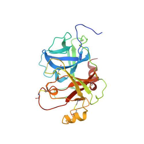
    1FDP

  • PubMed Abstract: 

    The crystal structure of profactor D, determined at 2.1 A resolution with an Rfree and an R-factor of 25.1 and 20.4%, respectively, displays highly flexible or disordered conformation for five regions: N-22, 71-76, 143-152, 187-193 and 215-223. A comparison with the structure of its mature serine protease, complement factor D, revealed major conformational changes in the similar regions. Comparisons with the zymogen-active enzyme pairs of chymotrypsinogen, trypsinogen and prethrombin-2 showed a similar distribution of the flexible regions. However, profactor D is the most flexible of the four, and its mature enzyme displays inactive, self-inhibited active site conformation. Examination of the surface properties of the N-terminus-binding pocket indicates that Ile16 may play the initial positioning role for the N-terminus, and Leu17 probably also helps in inducing the required conformational changes. This process, perhaps shared by most chymotrypsinogen-like zymogens, is followed by a factor D-unique step, the re-orientation of an external Arg218 to an internal position for salt-bridging with Asp189, leading to the generation of the self-inhibited factor D.


  • Organizational Affiliation

    Division of Clinical Immunology and Rheumatology, University of Alabama at Birmingham, Birmingham, AL 35294, USA.


Macromolecules
Find similar proteins by:  (by identity cutoff)  |  3D Structure
Entity ID: 1
MoleculeChains Sequence LengthOrganismDetailsImage
PROENZYME OF COMPLEMENT FACTOR D
A, B, C, D
235Homo sapiensMutation(s): 0 
EC: 3.4.21.46
UniProt & NIH Common Fund Data Resources
Find proteins for P00746 (Homo sapiens)
Explore P00746 
Go to UniProtKB:  P00746
PHAROS:  P00746
GTEx:  ENSG00000197766 
Entity Groups  
Sequence Clusters30% Identity50% Identity70% Identity90% Identity95% Identity100% Identity
UniProt GroupP00746
Sequence Annotations
Expand
  • Reference Sequence
Experimental Data & Validation

Experimental Data

  • Method: X-RAY DIFFRACTION
  • Resolution: 2.10 Å
  • R-Value Free: 0.251 
  • R-Value Work: 0.204 
  • R-Value Observed: 0.204 
  • Space Group: P 1 21 1
Unit Cell:
Length ( Å )Angle ( ˚ )
a = 65.05α = 90
b = 70.32β = 101.98
c = 86.3γ = 90
Software Package:
Software NamePurpose
DENZOdata reduction
SCALEPACKdata scaling
X-PLORmodel building
X-PLORrefinement
X-PLORphasing

Structure Validation

View Full Validation Report


Entry History 

Deposition Data

Revision History  (Full details and data files)

  • Version 1.0: 1999-12-03
    Type: Initial release
  • Version 1.1: 2008-04-26
    Changes: Version format compliance
  • Version 1.2: 2011-07-13
    Changes: Version format compliance
  • Version 1.3: 2011-12-07
    Changes: Database references
  • Version 1.4: 2023-08-09
    Changes: Data collection, Database references, Refinement description