1F8G

THE X-RAY STRUCTURE OF NICOTINAMIDE NUCLEOTIDE TRANSHYDROGENASE FROM RHODOSPIRILLUM RUBRUM COMPLEXED WITH NAD+


Experimental Data Snapshot

  • Method: X-RAY DIFFRACTION
  • Resolution: 2.00 Å
  • R-Value Free: 0.260 
  • R-Value Work: 0.210 

wwPDB Validation   3D Report Full Report


This is version 1.3 of the entry. See complete history


Literature

Protein-protein recognition, hydride transfer and proton pumping in the transhydrogenase complex.

Buckley, P.A.Baz Jackson, J.Schneider, T.White, S.A.Rice, D.W.Baker, P.J.

(2000) Structure 8: 809-815

  • DOI: https://doi.org/10.1016/s0969-2126(00)00171-4
  • Primary Citation of Related Structures:  
    1F8G

  • PubMed Abstract: 

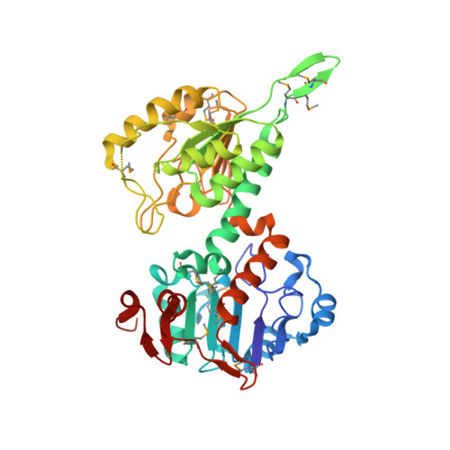
    Membrane-bound ion pumps are involved in metabolic regulation, osmoregulation, cell signalling, nerve transmission and energy transduction. How the ion electrochemical gradient interacts with the scalar chemistry and how the catalytic machinery is gated to ensure high coupling efficiency are fundamental to the mechanism of action of such pumps. Transhydrogenase is a conformationally coupled proton pump linking a proton gradient to the redox reaction between NAD(H) and NADP(H). The enzyme has three components; dI binds NAD(H), dII spans the membrane and dIII binds NADP(H).


  • Organizational Affiliation

    Department of Molecular Biology and Biotechnology, University of Sheffield, Western Bank, UK.


Macromolecules
Find similar proteins by:  (by identity cutoff)  |  3D Structure
Entity ID: 1
MoleculeChains Sequence LengthOrganismDetailsImage
NICOTINAMIDE NUCLEOTIDE TRANSHYDROGENASE
A, B, C, D
384Rhodospirillum rubrumMutation(s): 15 
EC: 1.6.1.1
UniProt
Find proteins for Q2RSB2 (Rhodospirillum rubrum (strain ATCC 11170 / ATH 1.1.1 / DSM 467 / LMG 4362 / NCIMB 8255 / S1))
Explore Q2RSB2 
Go to UniProtKB:  Q2RSB2
Entity Groups  
Sequence Clusters30% Identity50% Identity70% Identity90% Identity95% Identity100% Identity
UniProt GroupQ2RSB2
Sequence Annotations
Expand
  • Reference Sequence
Small Molecules
Modified Residues  1 Unique
IDChains TypeFormula2D DiagramParent
MSE
Query on MSE
A, B, C, D
L-PEPTIDE LINKINGC5 H11 N O2 SeMET
Experimental Data & Validation

Experimental Data

  • Method: X-RAY DIFFRACTION
  • Resolution: 2.00 Å
  • R-Value Free: 0.260 
  • R-Value Work: 0.210 
  • Space Group: P 1 21 1
Unit Cell:
Length ( Å )Angle ( ˚ )
a = 65.9α = 90
b = 116.6β = 104.22
c = 102γ = 90
Software Package:
Software NamePurpose
MLPHAREphasing
REFMACrefinement
DENZOdata reduction
SCALEPACKdata scaling

Structure Validation

View Full Validation Report


Entry History 

Deposition Data

Revision History  (Full details and data files)

  • Version 1.0: 2001-06-30
    Type: Initial release
  • Version 1.1: 2008-04-27
    Changes: Version format compliance
  • Version 1.2: 2011-07-13
    Changes: Version format compliance
  • Version 1.3: 2017-09-13
    Changes: Refinement description