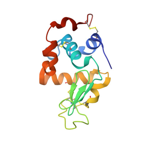
Crystal structures of apo- and holo-bovine alpha-lactalbumin at 2. 2-A resolution reveal an effect of calcium on inter-lobe interactions.
Chrysina, E.D., Brew, K., Acharya, K.R.(2000) J Biol Chem 275: 37021-37029
- PubMed: 10896943
- DOI: https://doi.org/10.1074/jbc.M004752200
- Primary Citation of Related Structures:
1F6R, 1F6S - PubMed Abstract:
High affinity binding of Ca(2+) to alpha-lactalbumin (LA) stabilizes the native structure and is required for the efficient generation of native protein with correct disulfide bonds from the reduced denatured state. A progressive increase in affinity of LA conformers for Ca(2+) as they develop increasingly native structures can account for the tendency of the apo form to assume a molten globule state and the large acceleration of folding by Ca(2+). To investigate the effect of calcium on structure of bovine LA, x-ray structures have been determined for crystals of the apo and holo forms at 2.2-A resolution. In both crystal forms, which were grown at high ionic strength, the protein is in a similar global native conformation consisting of alpha-helical and beta-subdomains separated by a cleft. Even though alternative cations and Ca(2+) liganding solvent molecules are absent, removal of Ca(2+) has only minor effects on the structure of the metal-binding site and a structural change was observed in the cleft on the opposite face of the molecule adjoining Tyr(103) of the helical lobe and Gln(54) of the beta-lobe. Changes include increased separation of the lobes, loss of a buried solvent molecule near the Ca(2+)-binding site, and the replacement of inter- and intra-lobe H-bonds of Tyr(103) by interactions with new immobilized water molecules. The more open cleft structure in the apo protein appears to be an effect of calcium binding transmitted via a change in orientation of helix H3 relative to the beta-lobe to the inter-lobe interface. Calcium is well known to promote the folding of LA. The results from the comparison of apo and holo structures of LA provide high resolution structural evidence that the acceleration of folding by Ca(2+) is mediated by an effect on interactions between the two subdomains.
Organizational Affiliation:
Department of Biology and Biochemistry, University of Bath, Claverton Down, Bath BA2 7AY, United Kingdom.