A ligand-reversible dimerization system for controlling protein-protein interactions.
Rollins, C.T., Rivera, V.M., Woolfson, D.N., Keenan, T., Hatada, M., Adams, S.E., Andrade, L.J., Yaeger, D., van Schravendijk, M.R., Holt, D.A., Gilman, M., Clackson, T.(2000) Proc Natl Acad Sci U S A 97: 7096-7101
- PubMed: 10852943
- DOI: https://doi.org/10.1073/pnas.100101997
- Primary Citation of Related Structures:
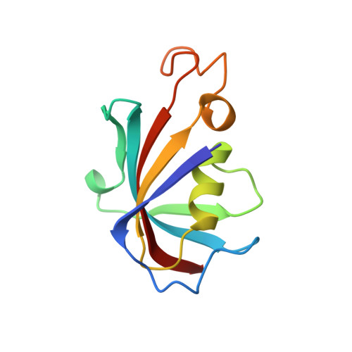
1EYM - PubMed Abstract:
Chemically induced dimerization provides a general way to gain control over intracellular processes. Typically, FK506-binding protein (FKBP) domains are fused to a signaling domain of interest, allowing crosslinking to be initiated by addition of a bivalent FKBP ligand. In the course of protein engineering studies on human FKBP, we discovered that a single point mutation in the ligand-binding site (Phe-36 --> Met) converts the normally monomeric protein into a ligand-reversible dimer. Two-hybrid, gel filtration, analytical ultracentrifugation, and x-ray crystallographic studies show that the mutant (F(M)) forms discrete homodimers with micromolar affinity that can be completely dissociated within minutes by addition of monomeric synthetic ligands. These unexpected properties form the basis for a "reverse dimerization" regulatory system involving F(M) fusion proteins, in which association is the ground state and addition of ligand abolishes interactions. We have used this strategy to rapidly and reversibly aggregate fusion proteins in different cellular compartments, and to provide an off switch for transcription. Reiterated F(M) domains should be generally useful as conditional aggregation domains (CADs) to control intracellular events where rapid, reversible dissolution of interactions is required. Our results also suggest that dimerization is a latent property of the FKBP fold: the crystal structure reveals a remarkably complementary interaction between the monomer binding sites, with only subtle changes in side-chain disposition accounting for the dramatic change in quaternary structure.
Organizational Affiliation:
ARIAD Gene Therapeutics, Inc., 26 Landsdowne Street, Cambridge, MA 02139, USA.