Protein crystallization by design: chymotrypsinogen without precipitants.
Pjura, P.E., Lenhoff, A.M., Leonard, S.A., Gittis, A.G.(2000) J Mol Biol 300: 235-239
- PubMed: 10873462
- DOI: https://doi.org/10.1006/jmbi.2000.3851
- Primary Citation of Related Structures:
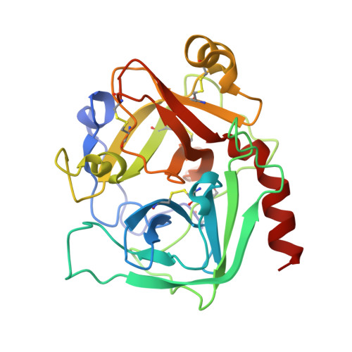
1EX3 - PubMed Abstract:
Protein crystals are usually obtained by an empirical approach based on extensive screening to identify suitable crystallization conditions. In contrast, we have used a systematic predictive procedure to produce data-quality crystals of bovine chymotrypsinogen A and used them to obtain a refined X-ray structure to 3 A resolution. Measurements of the osmotic second virial coefficient of chymotrypsinogen solutions were used to identify suitable solvent conditions, following which crystals were grown for approximately 30 hours by ultracentrifugal crystallization, without the use of any precipitants. Existing structures of chymotrypsinogen were obtained in solutions including 10-30 % ethanol, whereas simple buffered NaCl solutions were used here. The protein crystallized in the tetragonal space group P4(1)2(1)2, with one molecule per asymmetric unit. The quality of the refined map was very high throughout, with the main-chain atoms of all but four residues clearly defined and with nearly all side-chains also defined. Although only minor differences are seen compared to the structures previously reported, they indicate the possibility of structural changes due to the crystallization conditions used in those studies. Our results show that more systematic crystallization of proteins is possible, and that the procedure can expand the range of conditions under which crystals can be grown successfully and can make new crystal forms available.
Organizational Affiliation:
Center for Molecular and Engineering Thermodynamics, Department of Chemical Engineering, University of Delaware, Newark, DE 19716, USA.