1EK5

STRUCTURE OF HUMAN UDP-GALACTOSE 4-EPIMERASE IN COMPLEX WITH NAD+


Experimental Data Snapshot

  • Method: X-RAY DIFFRACTION
  • Resolution: 1.80 Å
  • R-Value Free: 0.223 
  • R-Value Work: 0.190 
  • R-Value Observed: 0.191 

wwPDB Validation   3D Report Full Report


Ligand Structure Quality Assessment 


This is version 1.4 of the entry. See complete history


Literature

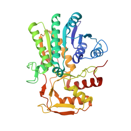
Crystallographic evidence for Tyr 157 functioning as the active site base in human UDP-galactose 4-epimerase.

Thoden, J.B.Wohlers, T.M.Fridovich-Keil, J.L.Holden, H.M.

(2000) Biochemistry 39: 5691-5701

  • DOI: https://doi.org/10.1021/bi000215l
  • Primary Citation of Related Structures:  
    1EK5, 1EK6

  • PubMed Abstract: 

    UDP-galactose 4-epimerase catalyzes the interconversion of UDP-glucose and UDP-galactose during normal galactose metabolism. In humans, deficiencies in this enzyme lead to the complex disorder referred to as epimerase-deficiency galactosemia. Here, we describe the high-resolution X-ray crystallographic structures of human epimerase in the resting state (i.e., with bound NAD(+)) and in a ternary complex with bound NADH and UDP-glucose. Those amino acid side chains responsible for anchoring the NAD(+) to the protein include Asp 33, Asn 37, Asp 66, Tyr 157, and Lys 161. The glucosyl group of the substrate is bound to the protein via the side-chain carboxamide groups of Asn 187 and Asn 207. Additionally, O(gamma) of Ser 132 and O(eta) of Tyr 157 lie within 2.4 and 3.1 A, respectively, of the 4'-hydroxyl group of the sugar. Comparison of the polypeptide chains for the resting enzyme and for the protein with bound NADH and UDP-glucose demonstrates that the major conformational changes which occur upon substrate binding are limited primarily to the regions defined by Glu 199 to Asp 240 and Gly 274 to Tyr 308. Additionally, this investigation reveals for the first time that a conserved tyrosine, namely Tyr 157, is in the proper position to interact directly with the 4'-hydroxyl group of the sugar substrate and to thus serve as the active-site base. A low barrier hydrogen bond between the 4'-hydroxyl group of the sugar and O(gamma) of Ser 132 facilitates proton transfer from the sugar 4'-hydroxyl group to O(eta) of Tyr 157.


  • Organizational Affiliation

    Department of Biochemistry, University of Wisconsin, Madison, Wisconsin 53705, USA.


Macromolecules
Find similar proteins by:  (by identity cutoff)  |  3D Structure
Entity ID: 1
MoleculeChains Sequence LengthOrganismDetailsImage
UDP-GALACTOSE 4-EPIMERASE348Homo sapiensMutation(s): 0 
EC: 5.1.3.2
UniProt & NIH Common Fund Data Resources
Find proteins for Q14376 (Homo sapiens)
Explore Q14376 
Go to UniProtKB:  Q14376
PHAROS:  Q14376
GTEx:  ENSG00000117308 
Entity Groups  
Sequence Clusters30% Identity50% Identity70% Identity90% Identity95% Identity100% Identity
UniProt GroupQ14376
Sequence Annotations
Expand
  • Reference Sequence
Small Molecules
Ligands 1 Unique
IDChains Name / Formula / InChI Key2D Diagram3D Interactions
NAD
Query on NAD

Download Ideal Coordinates CCD File 
B [auth A]NICOTINAMIDE-ADENINE-DINUCLEOTIDE
C21 H27 N7 O14 P2
BAWFJGJZGIEFAR-NNYOXOHSSA-N
Experimental Data & Validation

Experimental Data

  • Method: X-RAY DIFFRACTION
  • Resolution: 1.80 Å
  • R-Value Free: 0.223 
  • R-Value Work: 0.190 
  • R-Value Observed: 0.191 
  • Space Group: C 2 2 21
Unit Cell:
Length ( Å )Angle ( ˚ )
a = 50.3α = 90
b = 116.4β = 90
c = 130.4γ = 90
Software Package:
Software NamePurpose
TNTrefinement
FRAMBOdata collection
SAINTdata scaling
TNTphasing

Structure Validation

View Full Validation Report


Ligand Structure Quality Assessment 


Entry History 

Deposition Data

Revision History  (Full details and data files)

  • Version 1.0: 2000-05-17
    Type: Initial release
  • Version 1.1: 2008-04-27
    Changes: Version format compliance
  • Version 1.2: 2011-07-13
    Changes: Version format compliance
  • Version 1.3: 2017-10-04
    Changes: Refinement description
  • Version 1.4: 2024-02-07
    Changes: Data collection, Database references, Derived calculations