1EJ3

CRYSTAL STRUCTURE OF AEQUORIN


Experimental Data Snapshot

  • Method: X-RAY DIFFRACTION
  • Resolution: 2.30 Å
  • R-Value Free: 0.253 
  • R-Value Work: 0.219 
  • R-Value Observed: 0.221 

wwPDB Validation   3D Report Full Report


This is version 1.3 of the entry. See complete history


Literature

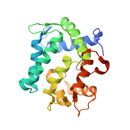
The crystal structure of the photoprotein aequorin at 2.3 A resolution.

Head, J.F.Inouye, S.Teranishi, K.Shimomura, O.

(2000) Nature 405: 372-376

  • DOI: https://doi.org/10.1038/35012659
  • Primary Citation of Related Structures:  
    1EJ3

  • PubMed Abstract: 

    Aequorin is a calcium-sensitive photoprotein originally obtained from the jellyfish Aequorea aequorea. Because it has a high sensitivity to calcium ions and is biologically harmless, aequorin is widely used as a probe to monitor intracellular levels of free calcium. The aequorin molecule contains four helix-loop-helix 'EF-hand' domains, of which three can bind calcium. The molecule also contains coelenterazine as its chromophoric ligand. When calcium is added, the protein complex decomposes into apoaequorin, coelenteramide and CO2, accompanied by the emission of light. Apoaequorin can be regenerated into active aequorin in the absence of calcium by incubation with coelenterazine, oxygen and a thiol agent. Cloning and expression of the complementary DNA for aequorin were first reported in 1985 (refs 2, 6), and growth of crystals of the recombinant protein has been described; however, techniques have only recently been developed to prepare recombinant aequorin of the highest purity, permitting a full crystallographic study. Here we report the structure of recombinant aequorin determined by X-ray crystallography. Aequorin is found to be a globular molecule containing a hydrophobic core cavity that accommodates the ligand coelenterazine-2-hydroperoxide. The structure shows protein components stabilizing the peroxide and suggests a mechanism by which calcium activation may occur.


  • Organizational Affiliation

    Department of Physiology, Boston University School of Medicine, Massachusetts 02118, USA. jfh@medxtal.bu.edu


Macromolecules
Find similar proteins by:  (by identity cutoff)  |  3D Structure
Entity ID: 1
MoleculeChains Sequence LengthOrganismDetailsImage
AEQUORIN
A, B
191Aequorea forskaleaMutation(s): 0 
UniProt
Find proteins for P02592 (Aequorea victoria)
Explore P02592 
Go to UniProtKB:  P02592
Entity Groups  
Sequence Clusters30% Identity50% Identity70% Identity90% Identity95% Identity100% Identity
UniProt GroupP02592
Sequence Annotations
Expand
  • Reference Sequence
Small Molecules
Ligands 1 Unique
IDChains Name / Formula / InChI Key2D Diagram3D Interactions
CZH
Query on CZH

Download Ideal Coordinates CCD File 
C [auth A],
D [auth B]
C2-HYDROPEROXY-COELENTERAZINE
C26 H21 N3 O5
HOSWCJDTHOAORT-SANMLTNESA-N
Experimental Data & Validation

Experimental Data

  • Method: X-RAY DIFFRACTION
  • Resolution: 2.30 Å
  • R-Value Free: 0.253 
  • R-Value Work: 0.219 
  • R-Value Observed: 0.221 
  • Space Group: P 43 21 2
Unit Cell:
Length ( Å )Angle ( ˚ )
a = 81.27α = 90
b = 81.27β = 90
c = 163.66γ = 90
Software Package:
Software NamePurpose
DENZOdata reduction
SCALEPACKdata scaling
SOLVEphasing
CNSrefinement

Structure Validation

View Full Validation Report


Entry History 

Deposition Data

Revision History  (Full details and data files)

  • Version 1.0: 2000-05-31
    Type: Initial release
  • Version 1.1: 2008-04-27
    Changes: Version format compliance
  • Version 1.2: 2011-07-13
    Changes: Version format compliance
  • Version 1.3: 2024-02-07
    Changes: Data collection, Database references, Derived calculations