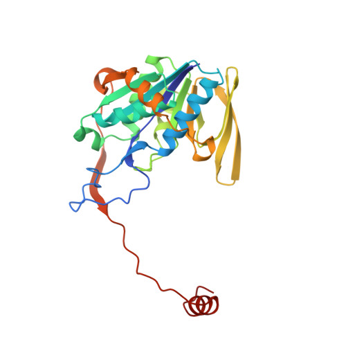
1EFV

THREE-DIMENSIONAL STRUCTURE OF HUMAN ELECTRON TRANSFER FLAVOPROTEIN TO 2.1 A RESOLUTION


Experimental Data Snapshot

  • Method: X-RAY DIFFRACTION
  • Resolution: 2.10 Å
  • R-Value Free: 0.222 
  • R-Value Work: 0.172 
  • R-Value Observed: 0.172 

wwPDB Validation   3D Report Full Report


This is version 1.3 of the entry. See complete history


Literature

Three-dimensional structure of human electron transfer flavoprotein to 2.1-A resolution.

Roberts, D.L.Frerman, F.E.Kim, J.J.

(1996) Proc Natl Acad Sci U S A 93: 14355-14360

  • DOI: https://doi.org/10.1073/pnas.93.25.14355
  • Primary Citation of Related Structures:  
    1EFV

  • PubMed Abstract: 

    Mammalian electron transfer flavoproteins (ETF) are heterodimers containing a single equivalent of flavin adenine dinucleotide (FAD). They function as electron shuttles between primary flavoprotein dehydrogenases involved in mitochondrial fatty acid and amino acid catabolism and the membrane-bound electron transfer flavoprotein ubiquinone oxidoreductase. The structure of human ETF solved to 2.1-A resolution reveals that the ETF molecule is comprised of three distinct domains: two domains are contributed by the alpha subunit and the third domain is made up entirely by the beta subunit. The N-terminal portion of the alpha subunit and the majority of the beta subunit have identical polypeptide folds, in the absence of any sequence homology. FAD lies in a cleft between the two subunits, with most of the FAD molecule residing in the C-terminal portion of the alpha subunit. Alignment of all the known sequences for the ETF alpha subunits together with the putative FixB gene product shows that the residues directly involved in FAD binding are conserved. A hydrogen bond is formed between the N5 of the FAD isoalloxazine ring and the hydroxyl side chain of alpha T266, suggesting why the pathogenic mutation, alpha T266M, affects ETF activity in patients with glutaric acidemia type II. Hydrogen bonds between the 4'-hydroxyl of the ribityl chain of FAD and N1 of the isoalloxazine ring, and between alpha H286 and the C2-carbonyl oxygen of the isoalloxazine ring, may play a role in the stabilization of the anionic semiquinone. With the known structure of medium chain acyl-CoA dehydrogenase, we hypothesize a possible structure for docking the two proteins.


  • Organizational Affiliation

    Department of Biochemistry, Medical College of Wisconsin, Milwaukee 53226, USA.


Macromolecules
Find similar proteins by:  (by identity cutoff)  |  3D Structure
Entity ID: 1
MoleculeChains Sequence LengthOrganismDetailsImage
ELECTRON TRANSFER FLAVOPROTEIN315Homo sapiensMutation(s): 0 
Membrane Entity: Yes 
UniProt & NIH Common Fund Data Resources
Find proteins for P13804 (Homo sapiens)
Explore P13804 
Go to UniProtKB:  P13804
PHAROS:  P13804
GTEx:  ENSG00000140374 
Entity Groups  
Sequence Clusters30% Identity50% Identity70% Identity90% Identity95% Identity100% Identity
UniProt GroupP13804
Sequence Annotations
Expand
  • Reference Sequence
Find similar proteins by:  (by identity cutoff)  |  3D Structure
Entity ID: 2
MoleculeChains Sequence LengthOrganismDetailsImage
ELECTRON TRANSFER FLAVOPROTEIN255Homo sapiensMutation(s): 0 
UniProt & NIH Common Fund Data Resources
Find proteins for P38117 (Homo sapiens)
Explore P38117 
Go to UniProtKB:  P38117
PHAROS:  P38117
GTEx:  ENSG00000105379 
Entity Groups  
Sequence Clusters30% Identity50% Identity70% Identity90% Identity95% Identity100% Identity
UniProt GroupP38117
Sequence Annotations
Expand
  • Reference Sequence
Small Molecules
Ligands 2 Unique
IDChains Name / Formula / InChI Key2D Diagram3D Interactions
FAD
Query on FAD

Download Ideal Coordinates CCD File 
C [auth A]FLAVIN-ADENINE DINUCLEOTIDE
C27 H33 N9 O15 P2
VWWQXMAJTJZDQX-UYBVJOGSSA-N
AMP
Query on AMP

Download Ideal Coordinates CCD File 
D [auth B]ADENOSINE MONOPHOSPHATE
C10 H14 N5 O7 P
UDMBCSSLTHHNCD-KQYNXXCUSA-N
Experimental Data & Validation

Experimental Data

  • Method: X-RAY DIFFRACTION
  • Resolution: 2.10 Å
  • R-Value Free: 0.222 
  • R-Value Work: 0.172 
  • R-Value Observed: 0.172 
  • Space Group: P 1 21 1
Unit Cell:
Length ( Å )Angle ( ˚ )
a = 47.437α = 90
b = 103.872β = 109.98
c = 63.696γ = 90
Software Package:
Software NamePurpose
HEAVYmodel building
PHASESphasing
X-PLORrefinement
DENZOdata reduction
SCALEPACKdata scaling
HEAVYphasing

Structure Validation

View Full Validation Report


Entry History 

Deposition Data

Revision History  (Full details and data files)

  • Version 1.0: 1997-12-03
    Type: Initial release
  • Version 1.1: 2008-03-24
    Changes: Version format compliance
  • Version 1.2: 2011-07-13
    Changes: Version format compliance
  • Version 1.3: 2024-02-07
    Changes: Data collection, Database references, Derived calculations