1ECZ

PROTEASE INHIBITOR ECOTIN


Experimental Data Snapshot

  • Method: X-RAY DIFFRACTION
  • Resolution: 2.68 Å
  • R-Value Free: 0.309 
  • R-Value Work: 0.195 
  • R-Value Observed: 0.195 

wwPDB Validation   3D Report Full Report


Ligand Structure Quality Assessment 


This is version 1.5 of the entry. See complete history


Literature

Crystal structure analyses of uncomplexed ecotin in two crystal forms: implications for its function and stability.

Shin, D.H.Song, H.K.Seong, I.S.Lee, C.S.Chung, C.H.Suh, S.W.

(1996) Protein Sci 5: 2236-2247

  • DOI: https://doi.org/10.1002/pro.5560051110
  • Primary Citation of Related Structures:  
    1ECY, 1ECZ

  • PubMed Abstract: 

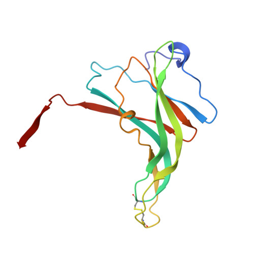
    Ecotin, a homodimeric protein composed of 142 residue subunits, is a novel serine protease inhibitor present in Escherichia coli. Its thermostability and acid stability, as well as broad specificity toward proteases, make it an interesting protein for structural characterization. Its structure in the uncomplexed state, determined for two different crystalline environments, allows a structural comparison of the free inhibitor with that in complex with trypsin. Although there is no gross structural rearrangement of ecotin when binding trypsin, the loops involved in binding trypsin show relatively large shifts in atomic positions. The inherent flexibility of the loops and the highly nonglobular shape are the two features essential for its inhibitory function. An insight into the understanding of the structural basis of thermostability and acid stability of ecotin is also provided by the present structure.


  • Organizational Affiliation

    Department of Chemistry, Seoul National University, Korea.


Macromolecules
Find similar proteins by:  (by identity cutoff)  |  3D Structure
Entity ID: 1
MoleculeChains Sequence LengthOrganismDetailsImage
ECOTIN
A, B
142Escherichia coliMutation(s): 0 
UniProt
Find proteins for P23827 (Escherichia coli (strain K12))
Explore P23827 
Go to UniProtKB:  P23827
Entity Groups  
Sequence Clusters30% Identity50% Identity70% Identity90% Identity95% Identity100% Identity
UniProt GroupP23827
Sequence Annotations
Expand
  • Reference Sequence
Small Molecules
Ligands 1 Unique
IDChains Name / Formula / InChI Key2D Diagram3D Interactions
BOG
Query on BOG

Download Ideal Coordinates CCD File 
C [auth A]
D [auth A]
E [auth A]
F [auth A]
G [auth A]
C [auth A],
D [auth A],
E [auth A],
F [auth A],
G [auth A],
H [auth A],
I [auth A],
J [auth A],
K [auth A],
L [auth A],
M [auth A],
N [auth B],
O [auth B],
P [auth B],
Q [auth B],
R [auth B],
S [auth B],
T [auth B],
U [auth B],
V [auth B],
W [auth B]
octyl beta-D-glucopyranoside
C14 H28 O6
HEGSGKPQLMEBJL-RKQHYHRCSA-N
Experimental Data & Validation

Experimental Data

  • Method: X-RAY DIFFRACTION
  • Resolution: 2.68 Å
  • R-Value Free: 0.309 
  • R-Value Work: 0.195 
  • R-Value Observed: 0.195 
  • Space Group: P 21 21 21
Unit Cell:
Length ( Å )Angle ( ˚ )
a = 39.22α = 90
b = 84.86β = 90
c = 98.74γ = 90
Software Package:
Software NamePurpose
MADNESdata collection
X-PLORmodel building
X-PLORrefinement
MADNESdata reduction
X-PLORphasing

Structure Validation

View Full Validation Report


Ligand Structure Quality Assessment 


Entry History 

Deposition Data

Revision History  (Full details and data files)

  • Version 1.0: 1997-02-12
    Type: Initial release
  • Version 1.1: 2008-03-24
    Changes: Version format compliance
  • Version 1.2: 2011-07-13
    Changes: Version format compliance
  • Version 1.3: 2018-04-18
    Changes: Advisory, Data collection, Other
  • Version 1.4: 2020-07-29
    Type: Remediation
    Reason: Carbohydrate remediation
    Changes: Advisory, Data collection, Derived calculations, Structure summary
  • Version 1.5: 2024-04-03
    Changes: Data collection, Database references, Refinement description, Structure summary