1ECF

ESCHERICHIA COLI GLUTAMINE PHOSPHORIBOSYLPYROPHOSPHATE (PRPP) AMIDOTRANSFERASE


Experimental Data Snapshot

  • Method: X-RAY DIFFRACTION
  • Resolution: 2.00 Å
  • R-Value Free: 0.230 
  • R-Value Work: 0.175 
  • R-Value Observed: 0.175 

wwPDB Validation   3D Report Full Report


This is version 1.4 of the entry. See complete history


Literature

Crystal structure of glutamine phosphoribosylpyrophosphate amidotransferase from Escherichia coli.

Muchmore, C.R.Krahn, J.M.Kim, J.H.Zalkin, H.Smith, J.L.

(1998) Protein Sci 7: 39-51

  • DOI: https://doi.org/10.1002/pro.5560070104
  • Primary Citation of Related Structures:  
    1ECF, 1ECJ

  • PubMed Abstract: 

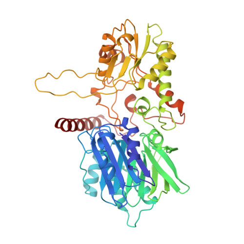
    Crystal structures of glutamine phosphoribosylpyrophosphate (PRPP) amidotransferase from Escherichia coli have been determined to 2.0-A resolution in the absence of ligands, and to 2.5-A resolution with the feedback inhibitor AMP bound to the PRPP catalytic site. Glutamine PRPP amidotransferase (GPATase) employs separate catalytic domains to abstract nitrogen from the amide of glutamine and to transfer nitrogen to the acceptor substrate PRPP. The unliganded and AMP-bound structures, which are essentially identical, are interpreted as the inhibited form of the enzyme because the two active sites are disconnected and the PRPP active site is solvent exposed. The structures were compared with a previously reported 3.0-A structure of the homologous Bacillus subtilis enzyme (Smith JL et al., 1994, Science 264:1427-1433). The comparison indicates a pattern of conservation of peptide structures involved with catalysis and variability in enzyme regulatory functions. Control of glutaminase activity, communication between the active sites, and regulation by feedback inhibitors are addressed differently by E. coli and B. subtilis GPATases. The E. coli enzyme is a prototype for the metal-free GPATases, whereas the B. subtilis enzyme represents the metal-containing enzymes. The structure of the E. coli enzyme suggests that a common ancestor of the two enzyme subfamilies may have included an Fe-S cluster.


  • Organizational Affiliation

    Department of Biological Sciences, Purdue University, West Lafayette, Indiana 47907, USA.


Macromolecules
Find similar proteins by:  (by identity cutoff)  |  3D Structure
Entity ID: 1
MoleculeChains Sequence LengthOrganismDetailsImage
GLUTAMINE PHOSPHORIBOSYLPYROPHOSPHATE AMIDOTRANSFERASE
A, B
504Escherichia coliMutation(s): 0 
Gene Names: PURF
EC: 2.4.2.14
UniProt
Find proteins for P0AG16 (Escherichia coli (strain K12))
Explore P0AG16 
Go to UniProtKB:  P0AG16
Entity Groups  
Sequence Clusters30% Identity50% Identity70% Identity90% Identity95% Identity100% Identity
UniProt GroupP0AG16
Sequence Annotations
Expand
  • Reference Sequence
Experimental Data & Validation

Experimental Data

  • Method: X-RAY DIFFRACTION
  • Resolution: 2.00 Å
  • R-Value Free: 0.230 
  • R-Value Work: 0.175 
  • R-Value Observed: 0.175 
  • Space Group: C 2 2 21
Unit Cell:
Length ( Å )Angle ( ˚ )
a = 116.9α = 90
b = 157.5β = 90
c = 106.3γ = 90
Software Package:
Software NamePurpose
SHARPphasing
X-PLORmodel building
X-PLORrefinement
HKLdata reduction
HKLdata scaling
SCALEPACKdata scaling
X-PLORphasing

Structure Validation

View Full Validation Report


Entry History 

Deposition Data

  • Released Date: 1996-11-08 
  • Deposition Author(s): Krahn, J.M.

Revision History  (Full details and data files)

  • Version 1.0: 1996-11-08
    Type: Initial release
  • Version 1.1: 2008-03-03
    Changes: Version format compliance
  • Version 1.2: 2011-07-13
    Changes: Derived calculations, Version format compliance
  • Version 1.3: 2011-11-16
    Changes: Atomic model
  • Version 1.4: 2023-08-09
    Changes: Advisory, Database references, Derived calculations, Refinement description