1EA3

Influenza virus M1 protein


Experimental Data Snapshot

  • Method: X-RAY DIFFRACTION
  • Resolution: 2.30 Å
  • R-Value Free: 0.313 
  • R-Value Work: 0.242 
  • R-Value Observed: 0.242 

wwPDB Validation   3D Report Full Report


This is version 2.1 of the entry. See complete history


Literature

Combined Results from Solution Studies on Intact Influenza Virus M1 Protein and from a New Crystal Form of its N-Terminal Domain Show that M1 is an Elongated Monomeric

Arzt, S.Baudin, F.Barge, A.Timmins, P.Burmeister, W.P.Ruigrok, R.W.

(2001) Virology 279: 439

  • DOI: https://doi.org/10.1006/viro.2000.0727
  • Primary Citation of Related Structures:  
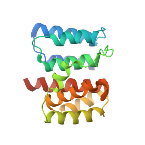
    1EA3

  • PubMed Abstract: 

    The amino-terminal domain of influenza A virus matrix protein (residues 1-164) was crystallized at pH 7 into a new crystal form in space group P1. This packing of the protein implies that M1(1-164) was monomeric in solution when it crystallized. Otherwise, the structure of the M1 fragment in the pH 7 crystals was the same as the monomers in crystals formed at pH 4 where crystal packing resulted in dimer formation [B. Sha and M. Luo, 1997, Nature Struct. Biol. 4, 239-244]. Analysis of intact M1 protein, the N-terminal domain, and the remaining C-terminal fragment (residues 165-252) in solution also showed that the N-terminal domain was monomeric with the same dimensions as determined from the crystal structure. Intact M1 protein was also monomeric but with an elongated shape due to the presence of the C-terminal part. Circular dichroism showed that the C-terminal part of M1 contained helical structure. A model for soluble M1 is presented, based on the assumption that the C-terminal domain is spherical, in which the N- and C-terminal domains are connected by a linker sequence which is available for proteolytic attack.


  • Organizational Affiliation

    European Synchrotron Radiation Facility, Forschungszentrum Jülich, Avenue des Martyrs, B.P. 220, Grenoble Cedex 09, F-38043, France.


Macromolecules
Find similar proteins by:  (by identity cutoff)  |  3D Structure
Entity ID: 1
MoleculeChains Sequence LengthOrganismDetailsImage
MATRIX PROTEIN M1
A, B
164Influenza A virusMutation(s): 0 
UniProt
Find proteins for P03485 (Influenza A virus (strain A/Puerto Rico/8/1934 H1N1))
Explore P03485 
Go to UniProtKB:  P03485
Entity Groups  
Sequence Clusters30% Identity50% Identity70% Identity90% Identity95% Identity100% Identity
UniProt GroupP03485
Sequence Annotations
Expand
  • Reference Sequence
Experimental Data & Validation

Experimental Data

  • Method: X-RAY DIFFRACTION
  • Resolution: 2.30 Å
  • R-Value Free: 0.313 
  • R-Value Work: 0.242 
  • R-Value Observed: 0.242 
  • Space Group: P 1
Unit Cell:
Length ( Å )Angle ( ˚ )
a = 36.86α = 77.1
b = 44.147β = 67.83
c = 47.692γ = 77.6
Software Package:
Software NamePurpose
MOSFLMdata reduction
SCALAdata scaling
AMoREphasing
EPMRphasing
CNSrefinement

Structure Validation

View Full Validation Report


Entry History 

Deposition Data

  • Released Date: 2001-04-26 
  • Deposition Author(s): Arzt, S.

Revision History  (Full details and data files)

  • Version 1.0: 2001-04-26
    Type: Initial release
  • Version 1.1: 2011-05-08
    Changes: Version format compliance
  • Version 1.2: 2011-07-13
    Changes: Version format compliance
  • Version 2.0: 2018-10-24
    Changes: Advisory, Atomic model, Data collection, Source and taxonomy
  • Version 2.1: 2023-12-13
    Changes: Advisory, Data collection, Database references, Other, Refinement description