1DZG

N135Q-S380C-ANTITHROMBIN-III


Experimental Data Snapshot

  • Method: X-RAY DIFFRACTION
  • Resolution: 2.80 Å
  • R-Value Free: 0.297 
  • R-Value Work: 0.228 

wwPDB Validation   3D Report Full Report


Ligand Structure Quality Assessment 


This is version 2.1 of the entry. See complete history


Literature

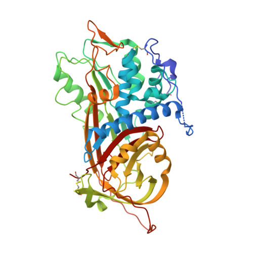
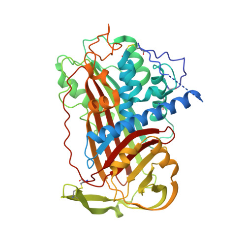
The Conformational Activation of Antithrombin. A 2. 85-A Structure of a Fluorescein Derivative Reveals an Electrostatic Link between the Hinge and Heparin Binding Regions.

Huntington, J.A.Mccoy, A.J.Belzar, K.J.Pei, X.Y.Gettins, P.G.W.Carrell, R.W.

(2000) J Biol Chem 275: 15377

  • DOI: https://doi.org/10.1074/jbc.275.20.15377
  • Primary Citation of Related Structures:  
    1DZG, 1DZH

  • PubMed Abstract: 

    Antithrombin is unique among the serpins in that it circulates in a native conformation that is kinetically inactive toward its target proteinase, factor Xa. Activation occurs upon binding of a specific pentasaccharide sequence found in heparin that results in a rearrangement of the reactive center loop removing constraints on the active center P1 residue. We determined the crystal structure of an activated antithrombin variant, N135Q S380C-fluorescein (P14-fluorescein), in order to see how full activation is achieved in the absence of heparin and how the structural effects of the substitution in the hinge region are translated to the heparin binding region. The crystal structure resembles native antithrombin except in the hinge and heparin binding regions. The absence of global conformational change allows for identification of specific interactions, centered on Glu(381) (P13), that are responsible for maintenance of the solution equilibrium between the native and activated forms and establishes the existence of an electrostatic link between the hinge region and the heparin binding region. A revised model for the mechanism of the allosteric activation of antithrombin is proposed.


  • Organizational Affiliation

    University of Cambridge, Department of Haematology, Wellcome Trust Centre for the Study of Molecular Mechanisms in Disease, Cambridge Institute for Medical Research, Hills Road, Cambridge CB2 2XY, United Kingdom. jah52@cam.ac.uk


Macromolecules
Find similar proteins by:  (by identity cutoff)  |  3D Structure
Entity ID: 1
MoleculeChains Sequence LengthOrganismDetailsImage
ANTITHROMBIN-IIIA [auth I]432Homo sapiensMutation(s): 2 
UniProt & NIH Common Fund Data Resources
Find proteins for P01008 (Homo sapiens)
Explore P01008 
Go to UniProtKB:  P01008
PHAROS:  P01008
GTEx:  ENSG00000117601 
Entity Groups  
Sequence Clusters30% Identity50% Identity70% Identity90% Identity95% Identity100% Identity
UniProt GroupP01008
Sequence Annotations
Expand
  • Reference Sequence
Find similar proteins by:  (by identity cutoff)  |  3D Structure
Entity ID: 2
MoleculeChains Sequence LengthOrganismDetailsImage
ANTITHROMBIN-IIIB [auth L]432Homo sapiensMutation(s): 0 
UniProt & NIH Common Fund Data Resources
Find proteins for P01008 (Homo sapiens)
Explore P01008 
Go to UniProtKB:  P01008
PHAROS:  P01008
GTEx:  ENSG00000117601 
Entity Groups  
Sequence Clusters30% Identity50% Identity70% Identity90% Identity95% Identity100% Identity
UniProt GroupP01008
Sequence Annotations
Expand
  • Reference Sequence
Oligosaccharides

Help

Entity ID: 3
MoleculeChains Length2D Diagram Glycosylation3D Interactions
2-acetamido-2-deoxy-beta-D-glucopyranose-(1-4)-2-acetamido-2-deoxy-beta-D-glucopyranoseC [auth A],
D [auth B],
E [auth C]
2N-Glycosylation
Glycosylation Resources
GlyTouCan:  G42666HT
GlyCosmos:  G42666HT
GlyGen:  G42666HT
Experimental Data & Validation

Experimental Data

  • Method: X-RAY DIFFRACTION
  • Resolution: 2.80 Å
  • R-Value Free: 0.297 
  • R-Value Work: 0.228 
  • Space Group: P 1 21 1
Unit Cell:
Length ( Å )Angle ( ˚ )
a = 68.61α = 90
b = 99.54β = 104.79
c = 88.69γ = 90
Software Package:
Software NamePurpose
REFMACrefinement
MOSFLMdata reduction
SCALAdata scaling
AMoREphasing

Structure Validation

View Full Validation Report


Ligand Structure Quality Assessment 


Entry History 

Deposition Data

Revision History  (Full details and data files)

  • Version 1.0: 2000-05-26
    Type: Initial release
  • Version 1.1: 2011-05-26
    Changes: Version format compliance
  • Version 1.2: 2011-07-13
    Changes: Version format compliance
  • Version 2.0: 2020-07-29
    Type: Remediation
    Reason: Carbohydrate remediation
    Changes: Atomic model, Data collection, Derived calculations, Other, Structure summary
  • Version 2.1: 2023-12-06
    Changes: Data collection, Database references, Refinement description, Structure summary