1DSZ

STRUCTURE OF THE RXR/RAR DNA-BINDING DOMAIN HETERODIMER IN COMPLEX WITH THE RETINOIC ACID RESPONSE ELEMENT DR1


Experimental Data Snapshot

  • Method: X-RAY DIFFRACTION
  • Resolution: 1.70 Å
  • R-Value Free: 0.267 
  • R-Value Work: 0.198 
  • R-Value Observed: 0.201 

wwPDB Validation   3D Report Full Report


This is version 1.3 of the entry. See complete history


Literature

Structure of the RXR-RAR DNA-binding complex on the retinoic acid response element DR1.

Rastinejad, F.Wagner, T.Zhao, Q.Khorasanizadeh, S.

(2000) EMBO J 19: 1045-1054

  • DOI: https://doi.org/10.1093/emboj/19.5.1045
  • Primary Citation of Related Structures:  
    1DSZ

  • PubMed Abstract: 

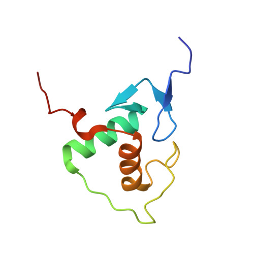
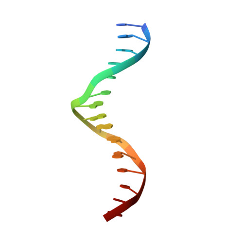
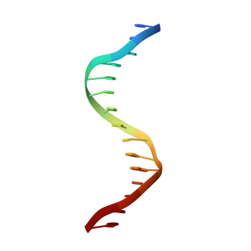
    The 9-cis retinoic acid receptor (retinoid X receptor, RXR) forms heterodimers with the all-trans retinoic acid receptor (RAR) and other nuclear receptors on DNA regulatory sites composed of tandem binding elements. We describe the 1.70 A resolution structure of the ternary complex of RXR and RAR DNA-binding regions in complex with the retinoic acid response element DR1. The receptors recognize identical half-sites through extensive base-specific contacts; however, RXR binds exclusively to the 3' site to form an asymmetric complex with the reverse polarity of other RXR heterodimers. The subunits associate in a strictly DNA-dependent manner using the T-box of RXR and the Zn-II region of RAR, both of which are reshaped in forming the complex. The protein-DNA contacts, the dimerization interface and the DNA curvature in the RXR-RAR complex are distinct from those of the RXR homodimer, which also binds DR1. Together, these structures illustrate how the nuclear receptor superfamily exploits conformational flexibility and locally induced structures to generate combinatorial transcription factors.


  • Organizational Affiliation

    Department of Pharmacology, University of Virginia, Charlottesville, VA 22908, USA,.


Macromolecules

Find similar proteins by:  (by identity cutoff)  |  3D Structure
Entity ID: 3
MoleculeChains Sequence LengthOrganismDetailsImage
RETINOIC ACID RECEPTOR ALPHAC [auth A]86Homo sapiensMutation(s): 0 
UniProt & NIH Common Fund Data Resources
Find proteins for P10276 (Homo sapiens)
Explore P10276 
Go to UniProtKB:  P10276
PHAROS:  P10276
GTEx:  ENSG00000131759 
Entity Groups  
Sequence Clusters30% Identity50% Identity70% Identity90% Identity95% Identity100% Identity
UniProt GroupP10276
Sequence Annotations
Expand
  • Reference Sequence
Find similar proteins by:  (by identity cutoff)  |  3D Structure
Entity ID: 4
MoleculeChains Sequence LengthOrganismDetailsImage
RETINOIC ACID RECEPTOR RXR-ALPHAD [auth B]85Homo sapiensMutation(s): 0 
UniProt & NIH Common Fund Data Resources
Find proteins for P19793 (Homo sapiens)
Explore P19793 
Go to UniProtKB:  P19793
PHAROS:  P19793
GTEx:  ENSG00000186350 
Entity Groups  
Sequence Clusters30% Identity50% Identity70% Identity90% Identity95% Identity100% Identity
UniProt GroupP19793
Sequence Annotations
Expand
  • Reference Sequence

Find similar nucleic acids by:  Sequence   |   3D Structure  

Entity ID: 1
MoleculeChains LengthOrganismImage
DNA (5'-D(*CP*AP*GP*GP*TP*CP*AP*AP*AP*GP*GP*TP*CP*AP*G)-3')A [auth C]15N/A
Sequence Annotations
Expand
  • Reference Sequence

Find similar nucleic acids by:  Sequence   |   3D Structure  

Entity ID: 2
MoleculeChains LengthOrganismImage
DNA (5'-D(*CP*TP*GP*AP*CP*CP*TP*TP*TP*GP*AP*CP*CP*TP*G)-3')B [auth D]15N/A
Sequence Annotations
Expand
  • Reference Sequence
Experimental Data & Validation

Experimental Data

  • Method: X-RAY DIFFRACTION
  • Resolution: 1.70 Å
  • R-Value Free: 0.267 
  • R-Value Work: 0.198 
  • R-Value Observed: 0.201 
  • Space Group: P 21 21 21
Unit Cell:
Length ( Å )Angle ( ˚ )
a = 80.66α = 90
b = 33.9β = 90
c = 101.86γ = 90
Software Package:
Software NamePurpose
AMoREphasing
CNSrefinement
DENZOdata reduction
SCALEPACKdata scaling

Structure Validation

View Full Validation Report


Entry History 

Deposition Data

Revision History  (Full details and data files)

  • Version 1.0: 2000-07-10
    Type: Initial release
  • Version 1.1: 2008-04-27
    Changes: Version format compliance
  • Version 1.2: 2011-07-13
    Changes: Version format compliance
  • Version 1.3: 2024-02-07
    Changes: Data collection, Database references, Derived calculations