1DOW

CRYSTAL STRUCTURE OF A CHIMERA OF BETA-CATENIN AND ALPHA-CATENIN


Experimental Data Snapshot

  • Method: X-RAY DIFFRACTION
  • Resolution: 1.80 Å
  • R-Value Free: 0.233 
  • R-Value Work: 0.200 
  • R-Value Observed: 0.200 

wwPDB Validation   3D Report Full Report


This is version 1.2 of the entry. See complete history


Literature

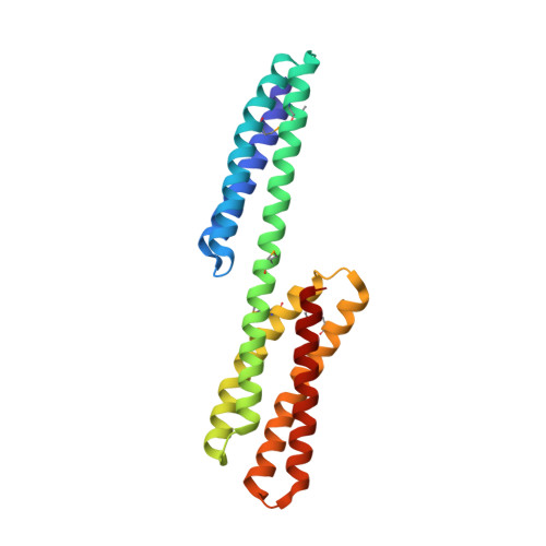
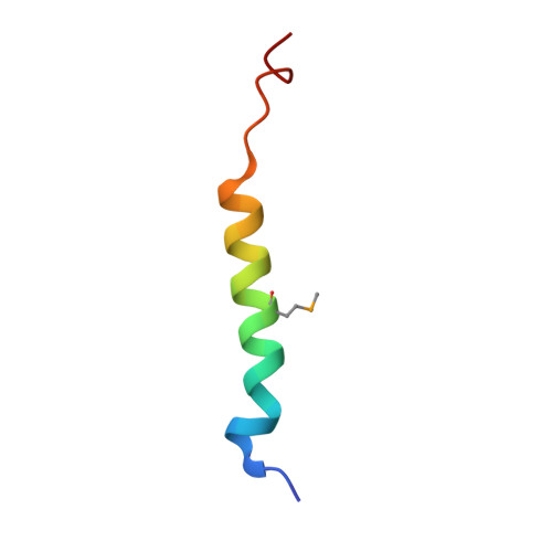
Structure of the dimerization and beta-catenin-binding region of alpha-catenin.

Pokutta, S.Weis, W.I.

(2000) Mol Cell 5: 533-543

  • DOI: https://doi.org/10.1016/s1097-2765(00)80447-5
  • Primary Citation of Related Structures:  
    1DOV, 1DOW

  • PubMed Abstract: 

    In adherens junctions, alpha-catenin links the cadherin-beta-catenin complex to the actin-based cytoskeleton. alpha-catenin is a homodimer in solution, but forms a 1:1 heterodimer with beta-catenin. The crystal structure of the alpha-catenin dimerization domain, residues 82-279, shows that alpha-catenin dimerizes through formation of a four-helix bundle in which two antiparallel helices are contributed by each protomer. A slightly larger fragment, comprising residues 57-264, binds to beta-catenin. A chimera consisting of the alpha-catenin-binding region of beta-catenin linked to the amino terminus of alpha-catenin 57-264 behaves as a monomer in solution, as expected, since beta-catenin binding disrupts the alpha-catenin dimer. The crystal structure of this chimera reveals the interaction between alpha- and beta-catenin, and provides a basis for understanding adherens junction assembly.


  • Organizational Affiliation

    Department of Structural Biology, Stanford University School of Medicine, California 94305, USA.


Macromolecules
Find similar proteins by:  (by identity cutoff)  |  3D Structure
Entity ID: 1
MoleculeChains Sequence LengthOrganismDetailsImage
ALPHA-CATENIN205Mus musculusMutation(s): 5 
UniProt
Find proteins for P26231 (Mus musculus)
Explore P26231 
Go to UniProtKB:  P26231
Entity Groups  
Sequence Clusters30% Identity50% Identity70% Identity90% Identity95% Identity100% Identity
UniProt GroupP26231
Sequence Annotations
Expand
  • Reference Sequence
Find similar proteins by:  (by identity cutoff)  |  3D Structure
Entity ID: 2
MoleculeChains Sequence LengthOrganismDetailsImage
BETA-CATENIN32Mus musculusMutation(s): 1 
UniProt & NIH Common Fund Data Resources
Find proteins for Q02248 (Mus musculus)
Explore Q02248 
Go to UniProtKB:  Q02248
IMPC:  MGI:88276
Entity Groups  
Sequence Clusters30% Identity50% Identity70% Identity90% Identity95% Identity100% Identity
UniProt GroupQ02248
Sequence Annotations
Expand
  • Reference Sequence
Small Molecules
Modified Residues  1 Unique
IDChains TypeFormula2D DiagramParent
MSE
Query on MSE
A
L-PEPTIDE LINKINGC5 H11 N O2 SeMET
Experimental Data & Validation

Experimental Data

  • Method: X-RAY DIFFRACTION
  • Resolution: 1.80 Å
  • R-Value Free: 0.233 
  • R-Value Work: 0.200 
  • R-Value Observed: 0.200 
  • Space Group: P 21 21 21
Unit Cell:
Length ( Å )Angle ( ˚ )
a = 53α = 90
b = 62.62β = 90
c = 63.66γ = 90
Software Package:
Software NamePurpose
CNSrefinement
DENZOdata reduction
SCALEPACKdata scaling
CNSphasing

Structure Validation

View Full Validation Report


Entry History 

Deposition Data

Revision History  (Full details and data files)

  • Version 1.0: 2000-07-12
    Type: Initial release
  • Version 1.1: 2008-04-27
    Changes: Version format compliance
  • Version 1.2: 2011-07-13
    Changes: Version format compliance