1D2N

D2 DOMAIN OF N-ETHYLMALEIMIDE-SENSITIVE FUSION PROTEIN


Experimental Data Snapshot

  • Method: X-RAY DIFFRACTION
  • Resolution: 1.75 Å
  • R-Value Free: 0.223 
  • R-Value Work: 0.199 
  • R-Value Observed: 0.199 

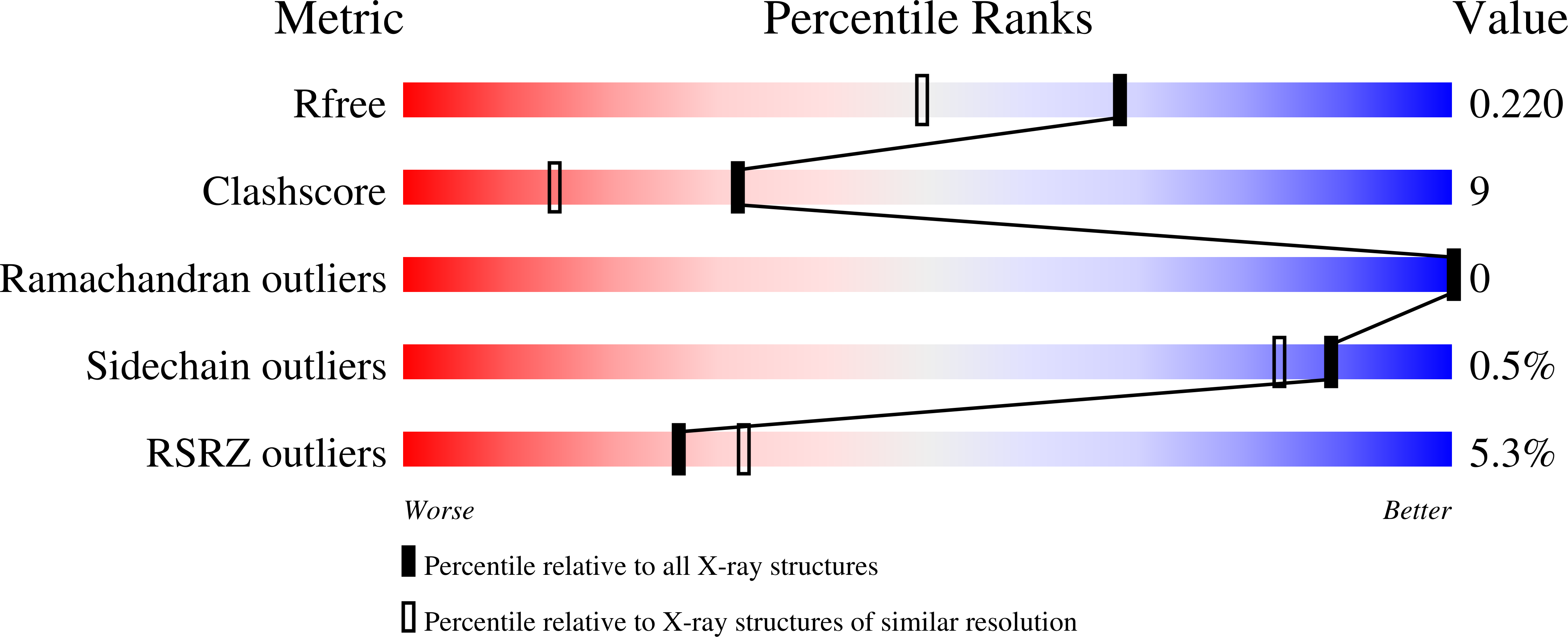
wwPDB Validation   3D Report Full Report


Ligand Structure Quality Assessment 


This is version 2.0 of the entry. See complete history


Literature

Crystal structure of the hexamerization domain of N-ethylmaleimide-sensitive fusion protein.

Lenzen, C.U.Steinmann, D.Whiteheart, S.W.Weis, W.I.

(1998) Cell 94: 525-536

  • DOI: https://doi.org/10.1016/s0092-8674(00)81593-7
  • Primary Citation of Related Structures:  
    1D2N

  • PubMed Abstract: 

    N-ethylmaleimide-sensitive fusion protein (NSF) is a cytosolic ATPase required for many intracellular vesicle fusion reactions. NSF consists of an amino-terminal region that interacts with other components of the vesicle trafficking machinery, followed by two homologous ATP-binding cassettes, designated D1 and D2, that possess essential ATPase and hexamerization activities, respectively. The crystal structure of D2 bound to Mg2+-AMPPNP has been determined at 1.75 A resolution. The structure consists of a nucleotide-binding and a helical domain, and it is unexpectedly similar to the first two domains of the clamp-loading subunit delta' of E. coli DNA polymerase III. The structure suggests several regions responsible for coupling of ATP hydrolysis to structural changes in full-length NSF.


  • Organizational Affiliation

    Department of Structural Biology, Stanford University School of Medicine, California 94305, USA.


Macromolecules
Find similar proteins by:  (by identity cutoff)  |  3D Structure
Entity ID: 1
MoleculeChains Sequence LengthOrganismDetailsImage
N-ETHYLMALEIMIDE-SENSITIVE FUSION PROTEIN272Cricetulus griseusMutation(s): 0 
UniProt
Find proteins for P18708 (Cricetulus griseus)
Explore P18708 
Go to UniProtKB:  P18708
Entity Groups  
Sequence Clusters30% Identity50% Identity70% Identity90% Identity95% Identity100% Identity
UniProt GroupP18708
Sequence Annotations
Expand
  • Reference Sequence
Experimental Data & Validation

Experimental Data

  • Method: X-RAY DIFFRACTION
  • Resolution: 1.75 Å
  • R-Value Free: 0.223 
  • R-Value Work: 0.199 
  • R-Value Observed: 0.199 
  • Space Group: P 6
Unit Cell:
Length ( Å )Angle ( ˚ )
a = 116.33α = 90
b = 116.33β = 90
c = 40.24γ = 120
Software Package:
Software NamePurpose
DMmodel building
MLPHAREphasing
CNSrefinement
DENZOdata reduction
SCALEPACKdata scaling
DMphasing

Structure Validation

View Full Validation Report


Ligand Structure Quality Assessment 


Entry History 

Deposition Data

Revision History  (Full details and data files)

  • Version 1.0: 1998-10-14
    Type: Initial release
  • Version 1.1: 2008-03-24
    Changes: Version format compliance
  • Version 1.2: 2011-07-13
    Changes: Derived calculations, Version format compliance
  • Version 2.0: 2024-02-07
    Changes: Atomic model, Data collection, Database references, Derived calculations