1CYY

CRYSTAL STRUCTURE OF THE 30 KDA FRAGMENT OF E. COLI DNA TOPOISOMERASE I. HEXAGONAL FORM


Experimental Data Snapshot

  • Method: X-RAY DIFFRACTION
  • Resolution: 2.15 Å
  • R-Value Free: 0.282 
  • R-Value Work: 0.229 
  • R-Value Observed: 0.234 

wwPDB Validation   3D Report Full Report


This is version 1.5 of the entry. See complete history


Literature

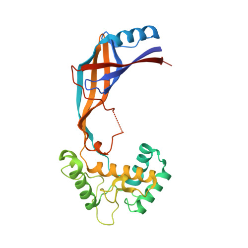
Conformational changes in E. coli DNA topoisomerase I.

Feinberg, H.Lima, C.D.Mondragon, A.

(1999) Nat Struct Biol 6: 918-922

  • DOI: https://doi.org/10.1038/13283
  • Primary Citation of Related Structures:  
    1CY9, 1CYY

  • PubMed Abstract: 

    DNA topoisomerases are the enzymes responsible for maintaining the topological states of DNA. In order to change the topology of DNA, topoisomerases pass one or two DNA strands through transient single or double strand breaks in the DNA phosphodiester backbone. It has been proposed that both type IA and type II enzymes change conformation dramatically during the reaction cycle in order to accomplish these transformations. In the case of Escherichia coli DNA topoisomerase I, it has been suggested that a 30 kDa fragment moves away from the rest of the protein to create an entrance into the central hole in the protein. Structures of the 30 kDa fragment reveal that indeed this fragment can change conformation significantly. The fragment is composed of two domains, and while the domains themselves remain largely unchanged, their relative arrangement can change dramatically.


  • Organizational Affiliation

    Department of Biochemistry, Molecular Biology and Cell Biology, Northwestern University, 2153 Sheridan Road, Evanston, Illinois 60208, USA.


Macromolecules
Find similar proteins by:  (by identity cutoff)  |  3D Structure
Entity ID: 1
MoleculeChains Sequence LengthOrganismDetailsImage
DNA TOPOISOMERASE I
A, B
264Escherichia coli K-12Mutation(s): 0 
EC: 5.99.1.2
UniProt
Find proteins for P06612 (Escherichia coli (strain K12))
Explore P06612 
Go to UniProtKB:  P06612
Entity Groups  
Sequence Clusters30% Identity50% Identity70% Identity90% Identity95% Identity100% Identity
UniProt GroupP06612
Sequence Annotations
Expand
  • Reference Sequence
Small Molecules
Ligands 1 Unique
IDChains Name / Formula / InChI Key2D Diagram3D Interactions
ZN
Query on ZN

Download Ideal Coordinates CCD File 
C [auth A]ZINC ION
Zn
PTFCDOFLOPIGGS-UHFFFAOYSA-N
Experimental Data & Validation

Experimental Data

  • Method: X-RAY DIFFRACTION
  • Resolution: 2.15 Å
  • R-Value Free: 0.282 
  • R-Value Work: 0.229 
  • R-Value Observed: 0.234 
  • Space Group: P 61 2 2
Unit Cell:
Length ( Å )Angle ( ˚ )
a = 113.85α = 90
b = 113.85β = 90
c = 233.87γ = 120
Software Package:
Software NamePurpose
CNSrefinement
REFMACrefinement
X-PLORrefinement
AMoREphasing
DENZOdata reduction
CCP4data scaling

Structure Validation

View Full Validation Report


Entry History 

Deposition Data

Revision History  (Full details and data files)

  • Version 1.0: 2000-03-08
    Type: Initial release
  • Version 1.1: 2008-04-27
    Changes: Version format compliance
  • Version 1.2: 2011-07-13
    Changes: Source and taxonomy, Version format compliance
  • Version 1.3: 2019-07-24
    Changes: Data collection, Refinement description
  • Version 1.4: 2019-08-14
    Changes: Data collection
  • Version 1.5: 2024-02-07
    Changes: Data collection, Database references, Derived calculations