1CVW

Crystal structure of active site-inhibited human coagulation factor VIIA (DES-GLA)


Experimental Data Snapshot

  • Method: X-RAY DIFFRACTION
  • Resolution: 2.28 Å
  • R-Value Free: 0.239 
  • R-Value Work: 0.205 
  • R-Value Observed: 0.205 

wwPDB Validation   3D Report Full Report


Ligand Structure Quality Assessment 


This is version 1.3 of the entry. See complete history


Literature

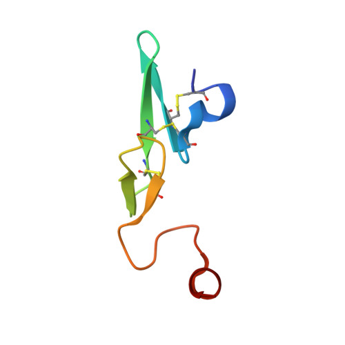
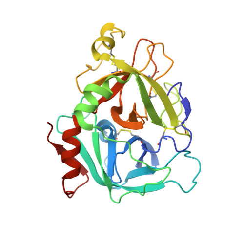
Crystal structure of active site-inhibited human coagulation factor VIIa (des-Gla).

Kemball-Cook, G.Johnson, D.J.Tuddenham, E.G.Harlos, K.

(1999) J Struct Biol 127: 213-223

  • DOI: https://doi.org/10.1006/jsbi.1999.4158
  • Primary Citation of Related Structures:  
    1CVW

  • PubMed Abstract: 

    Factor VIIa (FVIIa) is a crucial haemostatic protease consisting of four distinct domains termed the Gla, epidermal growth factor-1 (EGF-1), EGF-2, and protease domains (from N- to C-terminus). The crystal structure of human FVIIa inhibited at the active site with 1, 5-dansyl-Glu-Gly-Arg-chloromethyl ketone and lacking the Gla domain has been solved to a resolution of 2.28 A. The EGF-2 and protease domains were well resolved, whereas no electron density for the EGF-1 domain was observed, suggesting a flexible arrangement or disorder within the crystal. Superposition of the protease domain of the present structure with that previously resolved in the tissue factor (TF)/FVIIai complex revealed that although overall the domain structures are similar, the EGF-2 domain is rotated by 7.5 degrees relative to the protease domain on binding TF. A single cleavage in the protease domain was found, between Arg315 and Lys316 (chymotrypsin numbering 170C-170D) in a FVII-specific insertion loop: this cleavage appeared to be essential for crystallisation. Insertion of the heavy chain N-terminal Ile153 is essentially identical in the two structures, as is the geometry of the active site residues and the inhibitor C-terminal arginine residue. Some differences are seen in the cleaved loop, but changes in TF-contact residues are generally minor. This structure supports the hypothesis that TF binding enables spatial domain arrangements in the flexible FVIIa molecule necessary for procoagulant function and furthermore that active site occupancy induces FVIIa active conformation via N-terminal insertion.


  • Organizational Affiliation

    MRC Clinical Sciences Centre, Imperial College School of Medicine, Hammersmith Hospital, Du Cane Road, London, W12 0NN, United Kingdom. gkemball@hgmp.mrc.ac.uk


Macromolecules
Find similar proteins by:  (by identity cutoff)  |  3D Structure
Entity ID: 1
MoleculeChains Sequence LengthOrganismDetailsImage
COAGULATION FACTOR VIIA (LIGHT CHAIN) (DES-GLA)A [auth L]55Homo sapiensMutation(s): 0 
EC: 3.4.21.21
UniProt & NIH Common Fund Data Resources
Find proteins for P08709 (Homo sapiens)
Explore P08709 
Go to UniProtKB:  P08709
PHAROS:  P08709
GTEx:  ENSG00000057593 
Entity Groups  
Sequence Clusters30% Identity50% Identity70% Identity90% Identity95% Identity100% Identity
UniProt GroupP08709
Sequence Annotations
Expand
  • Reference Sequence
Find similar proteins by:  (by identity cutoff)  |  3D Structure
Entity ID: 2
MoleculeChains Sequence LengthOrganismDetailsImage
COAGULATION FACTOR VIIA (HEAVY CHAIN) (DES-GLA)B [auth H]254Homo sapiensMutation(s): 0 
EC: 3.4.21.21
UniProt & NIH Common Fund Data Resources
Find proteins for P08709 (Homo sapiens)
Explore P08709 
Go to UniProtKB:  P08709
PHAROS:  P08709
GTEx:  ENSG00000057593 
Entity Groups  
Sequence Clusters30% Identity50% Identity70% Identity90% Identity95% Identity100% Identity
UniProt GroupP08709
Sequence Annotations
Expand
  • Reference Sequence
Small Molecules
Ligands 2 Unique
IDChains Name / Formula / InChI Key2D Diagram3D Interactions
0GE
Query on 0GE

Download Ideal Coordinates CCD File 
C [auth H]N-{[5-(dimethylamino)naphthalen-1-yl]sulfonyl}-L-alpha-glutamyl-N-[(2S,3S)-6-carbamimidamido-1-chloro-2-hydroxyhexan-3-yl]glycinamide
C26 H38 Cl N7 O7 S
AKFRXQNHCCSRJN-IRFCIJBXSA-N
CA
Query on CA

Download Ideal Coordinates CCD File 
D [auth H]CALCIUM ION
Ca
BHPQYMZQTOCNFJ-UHFFFAOYSA-N
Biologically Interesting Molecules (External Reference) 1 Unique
Experimental Data & Validation

Experimental Data

  • Method: X-RAY DIFFRACTION
  • Resolution: 2.28 Å
  • R-Value Free: 0.239 
  • R-Value Work: 0.205 
  • R-Value Observed: 0.205 
  • Space Group: P 41 21 2
Unit Cell:
Length ( Å )Angle ( ˚ )
a = 94.85α = 90
b = 94.85β = 90
c = 114.3γ = 90
Software Package:
Software NamePurpose
DENZOdata reduction
SCALEPACKdata scaling
AMoREphasing
CNSrefinement

Structure Validation

View Full Validation Report


Ligand Structure Quality Assessment 


Entry History 

Deposition Data

Revision History  (Full details and data files)

  • Version 1.0: 1999-08-31
    Type: Initial release
  • Version 1.1: 2008-04-27
    Changes: Version format compliance
  • Version 1.2: 2011-07-13
    Changes: Atomic model, Database references, Derived calculations, Non-polymer description, Structure summary, Version format compliance
  • Version 1.3: 2013-02-27
    Changes: Non-polymer description