1CS6

N-TERMINAL FRAGMENT OF AXONIN-1 FROM CHICKEN


Experimental Data Snapshot

  • Method: X-RAY DIFFRACTION
  • Resolution: 1.80 Å
  • R-Value Free: 0.257 
  • R-Value Work: 0.226 
  • R-Value Observed: 0.226 

wwPDB Validation   3D Report Full Report


This is version 1.3 of the entry. See complete history


Literature

The crystal structure of the ligand binding module of axonin-1/TAG-1 suggests a zipper mechanism for neural cell adhesion.

Freigang, J.Proba, K.Leder, L.Diederichs, K.Sonderegger, P.Welte, W.

(2000) Cell 101: 425-433

  • DOI: https://doi.org/10.1016/s0092-8674(00)80852-1
  • Primary Citation of Related Structures:  
    1CS6

  • PubMed Abstract: 

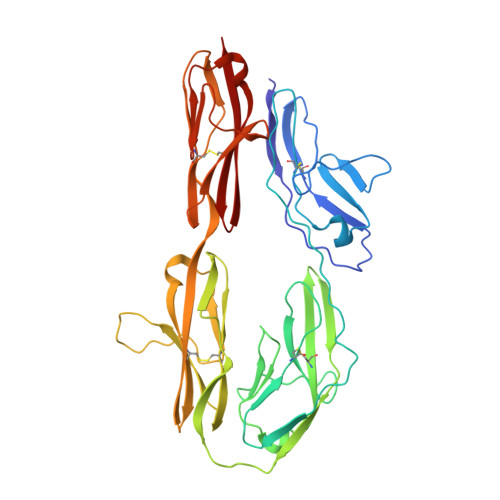
    We have determined the crystal structure of the ligand binding fragment of the neural cell adhesion molecule axonin-1/TAG-1 comprising the first four immunoglobulin (Ig) domains. The overall structure of axonin-1(Ig1-4) is U-shaped due to contacts between domains 1 and 4 and domains 2 and 3. In the crystals, these molecules are aligned in a string with adjacent molecules oriented in an anti-parallel fashion and their C termini perpendicular to the string. This arrangement suggests that cell adhesion by homophilic axonin-1 interaction occurs by the formation of a linear zipper-like array in which the axonin-1 molecules are alternately provided by the two apposed membranes. In accordance with this model, mutations in a loop critical for the formation of the zipper resulted in the loss of the homophilic binding capacity of axonin-1.


  • Organizational Affiliation

    Faculty of Biology, University of Konstanz, Germany.


Macromolecules
Find similar proteins by:  (by identity cutoff)  |  3D Structure
Entity ID: 1
MoleculeChains Sequence LengthOrganismDetailsImage
AXONIN-1382Gallus gallusMutation(s): 0 
UniProt
Find proteins for P28685 (Gallus gallus)
Explore P28685 
Go to UniProtKB:  P28685
Entity Groups  
Sequence Clusters30% Identity50% Identity70% Identity90% Identity95% Identity100% Identity
UniProt GroupP28685
Sequence Annotations
Expand
  • Reference Sequence
Small Molecules
Ligands 1 Unique
IDChains Name / Formula / InChI Key2D Diagram3D Interactions
GOL
Query on GOL

Download Ideal Coordinates CCD File 
B [auth A]GLYCEROL
C3 H8 O3
PEDCQBHIVMGVHV-UHFFFAOYSA-N
Experimental Data & Validation

Experimental Data

  • Method: X-RAY DIFFRACTION
  • Resolution: 1.80 Å
  • R-Value Free: 0.257 
  • R-Value Work: 0.226 
  • R-Value Observed: 0.226 
  • Space Group: P 1 21 1
Unit Cell:
Length ( Å )Angle ( ˚ )
a = 60.583α = 90
b = 45.54β = 96.14
c = 99.548γ = 90
Software Package:
Software NamePurpose
MAR345data collection
XDSdata reduction
SHARPphasing
CNSrefinement
XDSdata scaling

Structure Validation

View Full Validation Report


Entry History 

Deposition Data

Revision History  (Full details and data files)

  • Version 1.0: 2000-05-19
    Type: Initial release
  • Version 1.1: 2008-04-27
    Changes: Version format compliance
  • Version 1.2: 2011-07-13
    Changes: Non-polymer description, Version format compliance
  • Version 1.3: 2017-10-04
    Changes: Refinement description