1CRN

WATER STRUCTURE OF A HYDROPHOBIC PROTEIN AT ATOMIC RESOLUTION. PENTAGON RINGS OF WATER MOLECULES IN CRYSTALS OF CRAMBIN


Experimental Data Snapshot

  • Method: X-RAY DIFFRACTION
  • Resolution: 1.50 Å

wwPDB Validation   3D Report Full Report


This is version 1.4 of the entry. See complete history


Literature

Water structure of a hydrophobic protein at atomic resolution: Pentagon rings of water molecules in crystals of crambin.

Teeter, M.M.

(1984) Proc Natl Acad Sci U S A 81: 6014-6018

  • DOI: https://doi.org/10.1073/pnas.81.19.6014
  • Primary Citation of Related Structures:  
    1CRN

  • PubMed Abstract: 

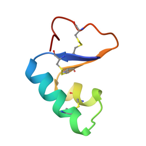
    The water structure has been analyzed for a model of the protein crambin refined against 0.945-A x-ray diffraction data. Crystals contain 32% solvent by volume, and 77% of the solvent molecules have been located-i.e., 2 ethanol molecules and 64 water molecules with 10-14 alternate positions. Many water oxygen atoms found form chains between polar groups on the surface of the protein. However, a cluster of pentagonal arrays made up of 16 water molecules sits at a hydrophobic, intermolecular cleft and forms a cap around the methyl group of leucine-18. Several waters in the cluster are hydrogen-bonded directly to the protein. Additional closed circular arrays, which include both protein atoms and other water oxygen atoms, form next to the central cluster. This water array stretches in the b lattice direction between groups of three ionic side chains.


  • Organizational Affiliation

    Department of Chemistry, Boston University, 685 Commonwealth Avenue, Boston, MA 02215.


Macromolecules
Find similar proteins by:  (by identity cutoff)  |  3D Structure
Entity ID: 1
MoleculeChains Sequence LengthOrganismDetailsImage
CRAMBIN46Crambe hispanica subsp. abyssinicaMutation(s): 0 
UniProt
Find proteins for P01542 (Crambe hispanica subsp. abyssinica)
Explore P01542 
Go to UniProtKB:  P01542
Entity Groups  
Sequence Clusters30% Identity50% Identity70% Identity90% Identity95% Identity100% Identity
UniProt GroupP01542
Sequence Annotations
Expand
  • Reference Sequence
Experimental Data & Validation

Experimental Data

  • Method: X-RAY DIFFRACTION
  • Resolution: 1.50 Å
  • Space Group: P 1 21 1
Unit Cell:
Length ( Å )Angle ( ˚ )
a = 40.96α = 90
b = 18.65β = 90.77
c = 22.52γ = 90
Software Package:
Software NamePurpose
PROLSQrefinement

Structure Validation

View Full Validation Report


Entry History 

Deposition Data

Revision History  (Full details and data files)

  • Version 1.0: 1981-07-28
    Type: Initial release
  • Version 1.1: 2008-03-24
    Changes: Version format compliance
  • Version 1.2: 2011-07-13
    Changes: Version format compliance
  • Version 1.3: 2012-07-11
    Changes: Other
  • Version 1.4: 2017-11-29
    Changes: Derived calculations, Other