1CKL

N-TERMINAL TWO DOMAINS OF HUMAN CD46 (MEMBRANE COFACTOR PROTEIN, MCP)


Experimental Data Snapshot

  • Method: X-RAY DIFFRACTION
  • Resolution: 3.10 Å
  • R-Value Free: 0.298 
  • R-Value Work: 0.244 
  • R-Value Observed: 0.244 

wwPDB Validation   3D Report Full Report


This is version 2.1 of the entry. See complete history


Literature

Crystal structure of two CD46 domains reveals an extended measles virus-binding surface.

Casasnovas, J.M.Larvie, M.Stehle, T.

(1999) EMBO J 18: 2911-2922

  • DOI: https://doi.org/10.1093/emboj/18.11.2911
  • Primary Citation of Related Structures:  
    1CKL

  • PubMed Abstract: 

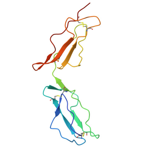
    Measles virus is a paramyxovirus which, like other members of the family such as respiratory syncytial virus, is a major cause of morbidity and mortality worldwide. The cell surface receptor for measles virus in humans is CD46, a complement cofactor. We report here the crystal structure at 3.1 A resolution of the measles virus-binding fragment of CD46. The structure reveals the architecture and spatial arrangement of two glycosylated short consensus repeats with a pronounced interdomain bend and some flexibility at the domain interface. Amino acids involved in measles virus binding define a large, glycan-free surface that extends from the top of the first to the bottom of the second repeat. The extended virus-binding surface of CD46 differs strikingly from those reported for the human virus receptor proteins CD4 and intercellular cell adhesion molecule-1 (ICAM-1), suggesting that the CD46 structure utilizes a novel mode of virus recognition. A highly hydrophobic and protruding loop at the base of the first repeat bears a critical virus-binding residue, thereby defining an important recognition epitope. Molecules that mimic the conformation of this loop potentially could be effective anti-viral agents by preventing binding of measles virus to CD46.


  • Organizational Affiliation

    Department of Biosciences at NOVUM, Karolinska Institute, 14157 Huddinge, Sweden.


Macromolecules
Find similar proteins by:  (by identity cutoff)  |  3D Structure
Entity ID: 1
MoleculeChains Sequence LengthOrganismDetailsImage
PROTEIN (CD46)
A, B, C, D, E
A, B, C, D, E, F
126Homo sapiensMutation(s): 0 
UniProt & NIH Common Fund Data Resources
Find proteins for P15529 (Homo sapiens)
Explore P15529 
Go to UniProtKB:  P15529
PHAROS:  P15529
GTEx:  ENSG00000117335 
Entity Groups  
Sequence Clusters30% Identity50% Identity70% Identity90% Identity95% Identity100% Identity
UniProt GroupP15529
Sequence Annotations
Expand
  • Reference Sequence
Oligosaccharides

Help

Entity ID: 2
MoleculeChains Length2D Diagram Glycosylation3D Interactions
alpha-D-mannopyranose-(1-2)-alpha-D-mannopyranose-(1-3)-alpha-D-mannopyranose-(1-6)-[alpha-D-mannopyranose-(1-2)-alpha-D-mannopyranose-(1-3)]beta-D-mannopyranose-(1-3)-2-acetamido-2-deoxy-beta-D-glucopyranose-(1-4)-2-acetamido-2-deoxy-beta-D-glucopyranose
G
8N-Glycosylation
Glycosylation Resources
GlyTouCan:  G63466HH
GlyCosmos:  G63466HH
GlyGen:  G63466HH
Entity ID: 3
MoleculeChains Length2D Diagram Glycosylation3D Interactions
alpha-D-mannopyranose-(1-3)-beta-D-mannopyranose-(1-3)-2-acetamido-2-deoxy-beta-D-glucopyranose-(1-4)-2-acetamido-2-deoxy-beta-D-glucopyranose
H
4N-Glycosylation
Glycosylation Resources
GlyTouCan:  G50737AO
GlyCosmos:  G50737AO
GlyGen:  G50737AO
Entity ID: 4
MoleculeChains Length2D Diagram Glycosylation3D Interactions
beta-D-mannopyranose-(1-4)-2-acetamido-2-deoxy-beta-D-glucopyranose-(1-4)-2-acetamido-2-deoxy-beta-D-glucopyranose
I, J, O
3N-Glycosylation
Glycosylation Resources
GlyTouCan:  G15407YE
GlyCosmos:  G15407YE
GlyGen:  G15407YE
Entity ID: 5
MoleculeChains Length2D Diagram Glycosylation3D Interactions
2-acetamido-2-deoxy-beta-D-glucopyranose-(1-4)-2-acetamido-2-deoxy-beta-D-glucopyranose
K, L, M
2N-Glycosylation
Glycosylation Resources
GlyTouCan:  G42666HT
GlyCosmos:  G42666HT
GlyGen:  G42666HT
Entity ID: 6
MoleculeChains Length2D Diagram Glycosylation3D Interactions
alpha-D-mannopyranose-(1-3)-[alpha-D-mannopyranose-(1-6)]beta-D-mannopyranose-(1-4)-2-acetamido-2-deoxy-beta-D-glucopyranose-(1-4)-2-acetamido-2-deoxy-beta-D-glucopyranose
N, R
5N-Glycosylation
Glycosylation Resources
GlyTouCan:  G22768VO
GlyCosmos:  G22768VO
GlyGen:  G22768VO
Entity ID: 7
MoleculeChains Length2D Diagram Glycosylation3D Interactions
alpha-D-mannopyranose-(1-2)-alpha-D-mannopyranose-(1-6)-[alpha-D-mannopyranose-(1-3)]alpha-D-mannopyranose-(1-6)-[alpha-D-mannopyranose-(1-3)]beta-D-mannopyranose-(1-4)-2-acetamido-2-deoxy-beta-D-glucopyranose-(1-4)-2-acetamido-2-deoxy-beta-D-glucopyranose
P
8N-Glycosylation
Glycosylation Resources
GlyTouCan:  G61846BY
GlyCosmos:  G61846BY
GlyGen:  G61846BY
Entity ID: 8
MoleculeChains Length2D Diagram Glycosylation3D Interactions
alpha-D-mannopyranose-(1-3)-[alpha-D-mannopyranose-(1-6)]alpha-D-mannopyranose-(1-6)-beta-D-mannopyranose-(1-4)-2-acetamido-2-deoxy-beta-D-glucopyranose-(1-4)-2-acetamido-2-deoxy-beta-D-glucopyranose
Q
6N-Glycosylation
Glycosylation Resources
GlyTouCan:  G94106MV
GlyCosmos:  G94106MV
GlyGen:  G94106MV
Small Molecules
Ligands 2 Unique
IDChains Name / Formula / InChI Key2D Diagram3D Interactions
CA
Query on CA

Download Ideal Coordinates CCD File 
BA [auth F]
S [auth A]
U [auth B]
W [auth C]
X [auth D]
BA [auth F],
S [auth A],
U [auth B],
W [auth C],
X [auth D],
Z [auth E]
CALCIUM ION
Ca
BHPQYMZQTOCNFJ-UHFFFAOYSA-N
CL
Query on CL

Download Ideal Coordinates CCD File 
AA [auth E],
CA [auth F],
T [auth A],
V [auth B],
Y [auth D]
CHLORIDE ION
Cl
VEXZGXHMUGYJMC-UHFFFAOYSA-M
Experimental Data & Validation

Experimental Data

  • Method: X-RAY DIFFRACTION
  • Resolution: 3.10 Å
  • R-Value Free: 0.298 
  • R-Value Work: 0.244 
  • R-Value Observed: 0.244 
  • Space Group: P 21 21 21
Unit Cell:
Length ( Å )Angle ( ˚ )
a = 74.823α = 90
b = 111.204β = 90
c = 136.986γ = 90
Software Package:
Software NamePurpose
DENZOdata reduction
SCALEPACKdata scaling
MLPHAREphasing
X-PLORrefinement

Structure Validation

View Full Validation Report


Entry History 

Deposition Data

Revision History  (Full details and data files)

  • Version 1.0: 1999-06-11
    Type: Initial release
  • Version 1.1: 2008-04-26
    Changes: Version format compliance
  • Version 1.2: 2011-07-13
    Changes: Non-polymer description, Version format compliance
  • Version 2.0: 2020-07-29
    Type: Remediation
    Reason: Carbohydrate remediation
    Changes: Advisory, Atomic model, Data collection, Derived calculations, Structure summary
  • Version 2.1: 2023-12-27
    Changes: Data collection, Database references, Structure summary