1CJM

HUMAN SULT1A3 WITH SULFATE BOUND


Experimental Data Snapshot

  • Method: X-RAY DIFFRACTION
  • Resolution: 2.40 Å
  • R-Value Free: 0.288 
  • R-Value Work: 0.276 

wwPDB Validation   3D Report Full Report


This is version 1.3 of the entry. See complete history


Literature

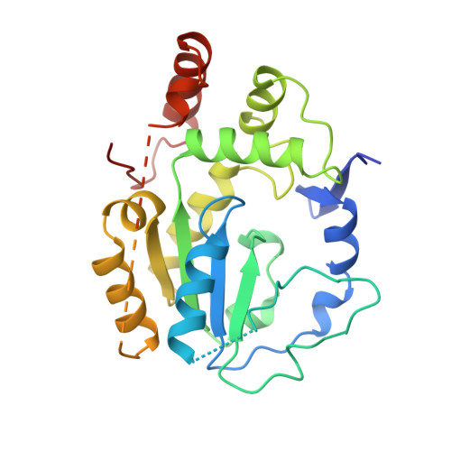
Crystal structure of human catecholamine sulfotransferase.

Bidwell, L.M.McManus, M.E.Gaedigk, A.Kakuta, Y.Negishi, M.Pedersen, L.Martin, J.L.

(1999) J Mol Biol 293: 521-530

  • DOI: https://doi.org/10.1006/jmbi.1999.3153
  • Primary Citation of Related Structures:  
    1CJM

  • PubMed Abstract: 

    Sulfonation, like phosphorylation, can modify the activity of a variety of biological molecules. The sulfotransferase enzymes sulfonate neurotransmitters, drugs, steroid hormones, dietary carcinogens and proteins. SULT1A3 specifically sulfonates catecholamines such as dopamine, adrenaline and noradrenaline. The crystal structure of SULT1A3 with a sulfate bound at the active site, has been determined at 2.4 A resolution. Although the core alpha/beta fold is like that of estrogen and heparan sulfotransferases, major differences occur in and around the active site. Most notably, several regions surrounding the active site, including a section of 40 residues, are disordered in SULT1A3. Regions that are topologically equivalent to the disordered parts of SULT1A3 are involved in substrate and cofactor binding in estrogen and heparan sulfotransferase. Flexibility in these regions suggests that ligand binding elicits a disorder-order transition in and around the active site of sulfotransferases and might contribute to the broad substrate specificity of these enzymes.


  • Organizational Affiliation

    Department of Physiology, University of Queensland, Brisbane, Queensland, 4072, Australia.


Macromolecules
Find similar proteins by:  (by identity cutoff)  |  3D Structure
Entity ID: 1
MoleculeChains Sequence LengthOrganismDetailsImage
PROTEIN (ARYL SULFOTRANSFERASE)295Homo sapiensMutation(s): 0 
UniProt & NIH Common Fund Data Resources
Find proteins for P0DMM9 (Homo sapiens)
Explore P0DMM9 
Go to UniProtKB:  P0DMM9
PHAROS:  P0DMM9
GTEx:  ENSG00000261052 
Entity Groups  
Sequence Clusters30% Identity50% Identity70% Identity90% Identity95% Identity100% Identity
UniProt GroupP0DMM9
Sequence Annotations
Expand
  • Reference Sequence
Small Molecules
Ligands 1 Unique
IDChains Name / Formula / InChI Key2D Diagram3D Interactions
SO4
Query on SO4

Download Ideal Coordinates CCD File 
B [auth A]SULFATE ION
O4 S
QAOWNCQODCNURD-UHFFFAOYSA-L
Experimental Data & Validation

Experimental Data

  • Method: X-RAY DIFFRACTION
  • Resolution: 2.40 Å
  • R-Value Free: 0.288 
  • R-Value Work: 0.276 
  • Space Group: P 32 2 1
Unit Cell:
Length ( Å )Angle ( ˚ )
a = 56.4α = 90
b = 56.4β = 90
c = 191.1γ = 120
Software Package:
Software NamePurpose
X-PLORmodel building
X-PLORrefinement
DENZOdata reduction
SCALEPACKdata scaling
X-PLORphasing

Structure Validation

View Full Validation Report


Entry History 

Deposition Data

Revision History  (Full details and data files)

  • Version 1.0: 1999-11-10
    Type: Initial release
  • Version 1.1: 2008-04-26
    Changes: Version format compliance
  • Version 1.2: 2011-07-13
    Changes: Version format compliance
  • Version 1.3: 2023-08-09
    Changes: Data collection, Database references, Derived calculations, Refinement description