1BOL

THE CRYSTAL STRUCTURE OF RIBONUCLEASE RH FROM RHIZOPUS NIVEUS AT 2.0 A RESOLUTION


Experimental Data Snapshot

  • Method: X-RAY DIFFRACTION
  • Resolution: 2.00 Å
  • R-Value Work: 0.185 
  • R-Value Observed: 0.185 

wwPDB Validation   3D Report Full Report


This is version 1.3 of the entry. See complete history


Literature

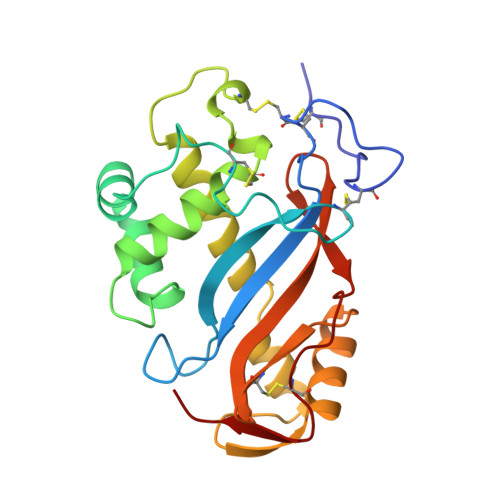
The crystal structure of ribonuclease Rh from Rhizopus niveus at 2.0 A resolution.

Kurihara, H.Nonaka, T.Mitsui, Y.Ohgi, K.Irie, M.Nakamura, K.T.

(1996) J Mol Biol 255: 310-320

  • DOI: https://doi.org/10.1006/jmbi.1996.0025
  • Primary Citation of Related Structures:  
    1BOL

  • PubMed Abstract: 

    The three-dimensional structure of ribonuclease Rh (RNase Rh), a new class of microbial ribonuclease from Rhizopus niveus, has been determined at 2.0 A resolution. The overall structure of RNase Rh is completely different from those of other previously studied RNases, such as RNase A from bovine pancreas and RNase T1 from Aspergillus oryzae. In the structure of RNase Rh, two histidine residues (His46 and His109) and one glutamic acid residue (Glu105), which were predicted to be critical to the activity from the chemical modification and mutagenesis experiments, are found to be located close together, constructing the active site. The indole ring of Trp49 plays an important role in preserving the active site structure by its stacking interactions with the imidazole ring of His 109, and by hydrogen bonding with the carboxyl group of Glu105. There exists a hydrophobic pocket around the active site, which contains the aromatic side-chain of Trp49 and Tyr57. The results of mutagenesis studies suggest that this pocket is the base binding site of the substrate.


  • Organizational Affiliation

    Department of BioEngineering, Nagaoka University of Technology, Japan.


Macromolecules
Find similar proteins by:  (by identity cutoff)  |  3D Structure
Entity ID: 1
MoleculeChains Sequence LengthOrganismDetailsImage
PROTEIN (RIBONUCLEASE RH)222Rhizopus niveusMutation(s): 0 
EC: 3.1.27.1
UniProt
Find proteins for P08056 (Rhizopus niveus)
Explore P08056 
Go to UniProtKB:  P08056
Entity Groups  
Sequence Clusters30% Identity50% Identity70% Identity90% Identity95% Identity100% Identity
UniProt GroupP08056
Sequence Annotations
Expand
  • Reference Sequence
Experimental Data & Validation

Experimental Data

  • Method: X-RAY DIFFRACTION
  • Resolution: 2.00 Å
  • R-Value Work: 0.185 
  • R-Value Observed: 0.185 
  • Space Group: P 21 21 21
Unit Cell:
Length ( Å )Angle ( ˚ )
a = 67.7α = 90
b = 72.5β = 90
c = 44.3γ = 90
Software Package:
Software NamePurpose
X-PLORrefinement

Structure Validation

View Full Validation Report


Entry History 

Deposition Data

Revision History  (Full details and data files)

  • Version 1.0: 1998-08-12
    Type: Initial release
  • Version 1.1: 2007-10-16
    Changes: Version format compliance
  • Version 1.2: 2011-07-13
    Changes: Version format compliance
  • Version 1.3: 2018-06-13
    Changes: Data collection