1BJF

CRYSTAL STRUCTURE OF RECOMBINANT BOVINE NEUROCALCIN DELTA AT 2.4 ANGSTROMS


Experimental Data Snapshot

  • Method: X-RAY DIFFRACTION
  • Resolution: 2.40 Å
  • R-Value Free: 0.308 
  • R-Value Work: 0.218 
  • R-Value Observed: 0.218 

wwPDB Validation   3D Report Full Report


This is version 1.3 of the entry. See complete history


Literature

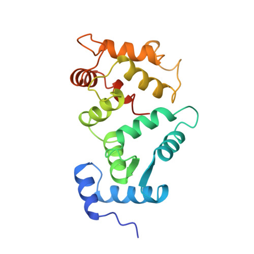
Crystal structure of recombinant bovine neurocalcin.

Vijay-Kumar, S.Kumar, V.D.

(1999) Nat Struct Biol 6: 80-88

  • DOI: https://doi.org/10.1038/4956
  • Primary Citation of Related Structures:  
    1BJF

  • PubMed Abstract: 

    The crystal structure of calcium-bound unmyristoylated bovine neurocalcin from Escherichia coli has been determined at 2.4 A resolution. The three-dimensional structure reveals a highly compact structure consisting of: (i) two pairs of calcium-binding EF-hands (EF1-EF2 and EF3-EF4); (ii) a calcium ion bound at EF2, EF3 and EF4 sites; and (iii) an EF1-hand that is disabled from calcium-binding due to a Cys-Pro sequence in the Ca2+-binding loop. The crystal structure of neurocalcin resembles photoreceptor recoverin in overall topology, however its EF2- and EF4-hands differ. Recently, neurocalcin in the calcium-bound state has been shown to stimulate mammalian rod outer segment membrane guanylate cyclase. A possible site for cyclase activity based on the three-dimensional structure is discussed.


  • Organizational Affiliation

    Department of Biochemistry, Fels Institute for Cancer Research and Molecular Biology, Temple University School of Medicine, Philadelphia, Pennsylvania 19140, USA.


Macromolecules
Find similar proteins by:  (by identity cutoff)  |  3D Structure
Entity ID: 1
MoleculeChains Sequence LengthOrganismDetailsImage
NEUROCALCIN DELTA
A, B
193Bos taurusMutation(s): 0 
UniProt
Find proteins for P61602 (Bos taurus)
Explore P61602 
Go to UniProtKB:  P61602
Entity Groups  
Sequence Clusters30% Identity50% Identity70% Identity90% Identity95% Identity100% Identity
UniProt GroupP61602
Sequence Annotations
Expand
  • Reference Sequence
Experimental Data & Validation

Experimental Data

  • Method: X-RAY DIFFRACTION
  • Resolution: 2.40 Å
  • R-Value Free: 0.308 
  • R-Value Work: 0.218 
  • R-Value Observed: 0.218 
  • Space Group: P 1 21 1
Unit Cell:
Length ( Å )Angle ( ˚ )
a = 42.68α = 90
b = 93.35β = 98.29
c = 50.625γ = 90
Software Package:
Software NamePurpose
X-PLORmodel building
X-PLORrefinement
R-AXISdata reduction
R-AXISdata scaling
X-PLORphasing

Structure Validation

View Full Validation Report


Entry History 

Deposition Data

Revision History  (Full details and data files)

  • Version 1.0: 1999-07-22
    Type: Initial release
  • Version 1.1: 2008-03-24
    Changes: Version format compliance
  • Version 1.2: 2011-07-13
    Changes: Version format compliance
  • Version 1.3: 2024-02-07
    Changes: Data collection, Database references, Derived calculations, Other