1BG5

CRYSTAL STRUCTURE OF THE ANKYRIN BINDING DOMAIN OF ALPHA-NA,K-ATPASE AS A FUSION PROTEIN WITH GLUTATHIONE S-TRANSFERASE


Experimental Data Snapshot

  • Method: X-RAY DIFFRACTION
  • Resolution: 2.60 Å
  • R-Value Free: 0.359 
  • R-Value Work: 0.193 
  • R-Value Observed: 0.193 

wwPDB Validation   3D Report Full Report


This is version 1.3 of the entry. See complete history


Literature

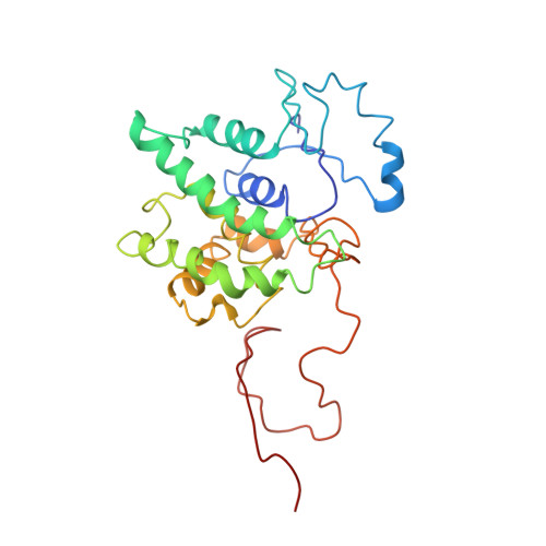
Structure of the ankyrin-binding domain of alpha-Na,K-ATPase.

Zhang, Z.Devarajan, P.Dorfman, A.L.Morrow, J.S.

(1998) J Biol Chem 273: 18681-18684

  • DOI: https://doi.org/10.1074/jbc.273.30.18681
  • Primary Citation of Related Structures:  
    1BG5

  • PubMed Abstract: 

    The ankyrin 33-residue repeating motif, an L-shaped structure with protruding beta-hairpin tips, mediates specific macromolecular interactions with cytoskeletal, membrane, and regulatory proteins. The association between ankyrin and alpha-Na,K-ATPase, a ubiquitous membrane protein critical to vectorial transport of ions and nutrients, is required to assemble and stabilize Na,K-ATPase at the plasma membrane. alpha-Na,K-ATPase binds both red cell ankyrin (AnkR, a product of the ANK1 gene) and Madin-Darby canine kidney cell ankyrin (AnkG, a product of the ANK3 gene) utilizing residues 142-166 (SYYQEAKSSKIMESFK NMVPQQALV) in its second cytoplasmic domain. Fusion peptides of glutathione S-transferase incorporating these 25 amino acids bind specifically to purified ankyrin (Kd = 118 +/- 50 nM). The three-dimensional structure (2.6 A) of this minimal ankyrin-binding motif, crystallized as the fusion protein, reveals a 7-residue loop with one charged hydrophilic face capping a double beta-strand. Comparison with ankyrin-binding sequences in p53, CD44, neurofascin/L1, and the inositol 1,4,5-trisphosphate receptor suggests that the valency and specificity of ankyrin binding is achieved by the interaction of 5-7-residue surface loops with the beta-hairpin tips of multiple ankyrin repeat units.


  • Organizational Affiliation

    Department of Pathology, Yale University School of Medicine, New Haven, Connecticut 06510, USA.


Macromolecules
Find similar proteins by:  (by identity cutoff)  |  3D Structure
Entity ID: 1
MoleculeChains Sequence LengthOrganismDetailsImage
FUSION PROTEIN OF ALPHA-NA,K-ATPASE WITH GLUTATHIONE S-TRANSFERASE254Rattus norvegicusMutation(s): 0 
UniProt
Find proteins for P08515 (Schistosoma japonicum)
Explore P08515 
Go to UniProtKB:  P08515
Entity Groups  
Sequence Clusters30% Identity50% Identity70% Identity90% Identity95% Identity100% Identity
UniProt GroupP08515
Sequence Annotations
Expand
  • Reference Sequence
Experimental Data & Validation

Experimental Data

  • Method: X-RAY DIFFRACTION
  • Resolution: 2.60 Å
  • R-Value Free: 0.359 
  • R-Value Work: 0.193 
  • R-Value Observed: 0.193 
  • Space Group: P 43 21 2
Unit Cell:
Length ( Å )Angle ( ˚ )
a = 92.17α = 90
b = 92.17β = 90
c = 57.57γ = 90
Software Package:
Software NamePurpose
AMoREphasing
X-PLORrefinement
DENZOdata reduction
SCALEPACKdata scaling

Structure Validation

View Full Validation Report


Entry History 

Deposition Data

Revision History  (Full details and data files)

  • Version 1.0: 1999-01-13
    Type: Initial release
  • Version 1.1: 2008-03-24
    Changes: Version format compliance
  • Version 1.2: 2011-07-13
    Changes: Version format compliance
  • Version 1.3: 2023-08-02
    Changes: Database references, Refinement description