1BBR

THE STRUCTURE OF RESIDUES 7-16 OF THE A ALPHA CHAIN OF HUMAN FIBRINOGEN BOUND TO BOVINE THROMBIN AT 2.3 ANGSTROMS RESOLUTION


Experimental Data Snapshot

  • Method: X-RAY DIFFRACTION
  • Resolution: 2.30 Å
  • R-Value Observed: 0.167 

wwPDB Validation   3D Report Full Report


This is version 1.2 of the entry. See complete history


Literature

The structure of residues 7-16 of the A alpha-chain of human fibrinogen bound to bovine thrombin at 2.3-A resolution.

Martin, P.D.Robertson, W.Turk, D.Huber, R.Bode, W.Edwards, B.F.

(1992) J Biol Chem 267: 7911-7920

  • Primary Citation of Related Structures:  
    1BBR

  • PubMed Abstract: 

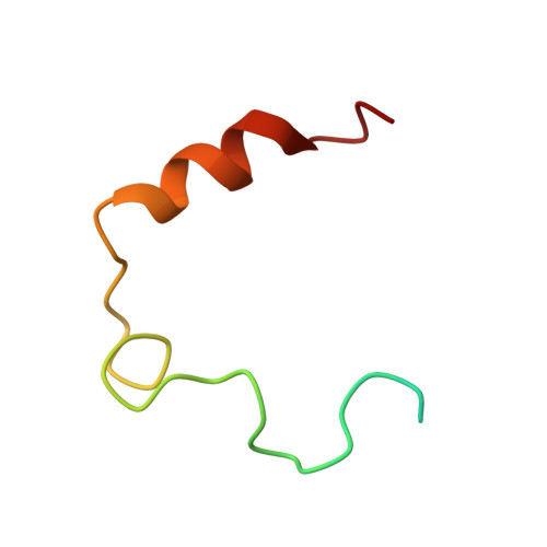
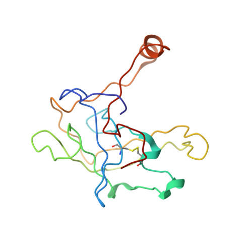
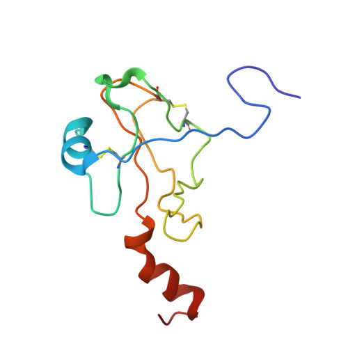
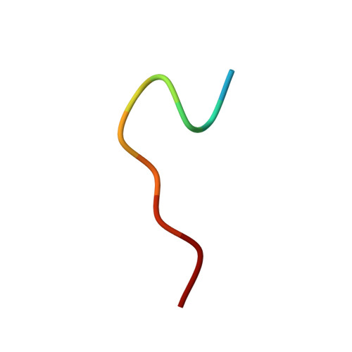
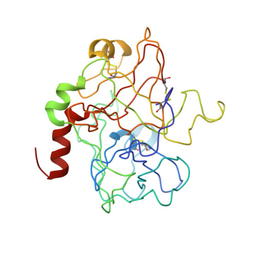
    The tetradecapeptide Ac-D-F-L-A-E-G-G-G-V-R-G-P-R-V-OMe, which mimics residues 7f-20f of the A alpha-chain of human fibrinogen, has been co-crystallized with bovine thrombin from ammonium sulfate solutions in space group P2(1) with unit cell dimensions of a = 83.0 A, b = 89.4 A, c = 99.3 A, and beta = 106.6 degrees. Three crystallographically independent complexes were located in the asymmetric unit by molecular replacement using the native bovine thrombin structure as a model. The standard crystallographic R-factor is 0.167 at 2.3-A resolution. Excellent electron density could be traced for the decapeptide, beginning with Asp-7f and ending with Arg-16f in the active site of thrombin; the remaining 4 residues, which have been cleaved from the tetradecapeptide at the Arg-16f/Gly-17f bond, are not seen. Residues 7f-11f at the NH2 terminus of the peptide form a single turn of alpha-helix that is connected by Gly-12f, which has a positive phi angle, to an extended chain containing residues 13f-16f. The major specific interactions between the peptide and thrombin are 1) a hydrophobic cage formed by residues Tyr-60A, Trp-60D, Leu-99, Ile-174, Trp-215, Leu-9f, Gly-13f, and Val-15f that surrounds Phe-8f; 2) a hydrogen bond linking Phe-8f NH to Lys-97 O;3) a salt link between Glu-11f and Arg-173; 4) two antiparallel beta-sheet hydrogen bonds between Gly-14f and Gly-216; and 5) the insertion of Arg-16f into the specificity pocket. Binding of the peptide is accompanied by a considerable shift in two of the loops near the active site relative to human D-phenyl-L-prolyl-L-arginyl chloromethyl ketone (PPACK)-thrombin.


  • Organizational Affiliation

    Department of Biochemistry, Wayne State University, Detroit, Michigan 48201.


Macromolecules
Find similar proteins by:  (by identity cutoff)  |  3D Structure
Entity ID: 1
MoleculeChains Sequence LengthOrganismDetailsImage
EPSILON-THROMBINA [auth L],
E [auth J],
H [auth M]
49Bos taurusMutation(s): 0 
EC: 3.4.21.5
UniProt
Find proteins for P00735 (Bos taurus)
Explore P00735 
Go to UniProtKB:  P00735
Entity Groups  
Sequence Clusters30% Identity50% Identity70% Identity90% Identity95% Identity100% Identity
UniProt GroupP00735
Sequence Annotations
Expand
  • Reference Sequence
Find similar proteins by:  (by identity cutoff)  |  3D Structure
Entity ID: 2
MoleculeChains Sequence LengthOrganismDetailsImage
EPSILON-THROMBINB [auth H]150Bos taurusMutation(s): 0 
EC: 3.4.21.5
UniProt
Find proteins for P00735 (Bos taurus)
Explore P00735 
Go to UniProtKB:  P00735
Entity Groups  
Sequence Clusters30% Identity50% Identity70% Identity90% Identity95% Identity100% Identity
UniProt GroupP00735
Sequence Annotations
Expand
  • Reference Sequence
Find similar proteins by:  (by identity cutoff)  |  3D Structure
Entity ID: 3
MoleculeChains Sequence LengthOrganismDetailsImage
EPSILON-THROMBINC [auth E]109Bos taurusMutation(s): 0 
EC: 3.4.21.5
UniProt
Find proteins for P00735 (Bos taurus)
Explore P00735 
Go to UniProtKB:  P00735
Entity Groups  
Sequence Clusters30% Identity50% Identity70% Identity90% Identity95% Identity100% Identity
UniProt GroupP00735
Sequence Annotations
Expand
  • Reference Sequence

Find similar proteins by:  Sequence   |   3D Structure  

Entity ID: 4
MoleculeChains Sequence LengthOrganismDetailsImage
FIBRINOGEN ALPHA/ALPHA-E CHAIN PRECURSORD [auth F],
G,
J [auth I]
11Homo sapiensMutation(s): 0 
UniProt & NIH Common Fund Data Resources
Find proteins for P02671 (Homo sapiens)
Explore P02671 
Go to UniProtKB:  P02671
PHAROS:  P02671
GTEx:  ENSG00000171560 
Entity Groups  
Sequence Clusters30% Identity50% Identity70% Identity90% Identity95% Identity100% Identity
UniProt GroupP02671
Sequence Annotations
Expand
  • Reference Sequence
Find similar proteins by:  (by identity cutoff)  |  3D Structure
Entity ID: 5
MoleculeChains Sequence LengthOrganismDetailsImage
EPSILON-THROMBINF [auth K],
I [auth N]
259Bos taurusMutation(s): 0 
EC: 3.4.21.5
UniProt
Find proteins for P00735 (Bos taurus)
Explore P00735 
Go to UniProtKB:  P00735
Entity Groups  
Sequence Clusters30% Identity50% Identity70% Identity90% Identity95% Identity100% Identity
UniProt GroupP00735
Sequence Annotations
Expand
  • Reference Sequence
Experimental Data & Validation

Experimental Data

  • Method: X-RAY DIFFRACTION
  • Resolution: 2.30 Å
  • R-Value Observed: 0.167 
  • Space Group: P 1 21 1
Unit Cell:
Length ( Å )Angle ( ˚ )
a = 83.02α = 90
b = 89.41β = 106.64
c = 99.3γ = 90
Software Package:
Software NamePurpose
PROLSQrefinement

Structure Validation

View Full Validation Report


Entry History 

Deposition Data

Revision History  (Full details and data files)

  • Version 1.0: 1994-01-31
    Type: Initial release
  • Version 1.1: 2008-03-24
    Changes: Version format compliance
  • Version 1.2: 2011-07-13
    Changes: Version format compliance