1AZZ

FIDDLER CRAB COLLAGENASE COMPLEXED TO ECOTIN


Experimental Data Snapshot

  • Method: X-RAY DIFFRACTION
  • Resolution: 2.30 Å
  • R-Value Free: 0.238 
  • R-Value Work: 0.189 
  • R-Value Observed: 0.189 

wwPDB Validation   3D Report Full Report


This is version 1.2 of the entry. See complete history


Literature

Crystal structure of an ecotin-collagenase complex suggests a model for recognition and cleavage of the collagen triple helix.

Perona, J.J.Tsu, C.A.Craik, C.S.Fletterick, R.J.

(1997) Biochemistry 36: 5381-5392

  • DOI: https://doi.org/10.1021/bi9617522
  • Primary Citation of Related Structures:  
    1AZZ

  • PubMed Abstract: 

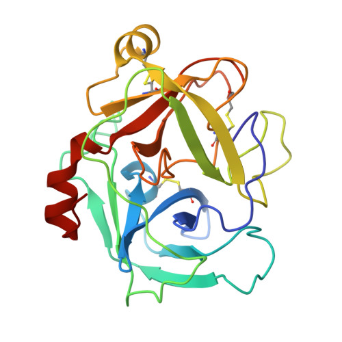
    The crystal structure of fiddler crab collagenase complexed with the dimeric serine protease inhibitor ecotin at 2.5 A resolution reveals an extended cleft providing binding sites for at least 11 contiguous substrate residues. Comparison of the positions of nine intermolecular main chain hydrogen bonding interactions in the cleft, with the known sequences at the cleavage site of type I collagen, suggests that the protease binding loop of ecotin adopts a conformation mimicking that of the cleaved strand of collagen. A well-defined groove extending across the binding surface of the enzyme readily accommodates the two other polypeptide chains of the triple-helical substrate. These observations permit construction of a detailed molecular model for collagen recognition and cleavage by this invertebrate serine protease. Ecotin undergoes a pronounced internal structural rearrangement which permits binding in the observed conformation. The capacity for such rearrangement appears to be a key determinant of its ability to inhibit a wide range of serine proteases.


  • Organizational Affiliation

    Department of Pharmaceutical Chemistry, University of California, San Francisco 94143-0446, USA.


Macromolecules
Find similar proteins by:  (by identity cutoff)  |  3D Structure
Entity ID: 1
MoleculeChains Sequence LengthOrganismDetailsImage
COLLAGENASE
A, B
226Leptuca pugilatorMutation(s): 0 
EC: 3.4.21.32
UniProt
Find proteins for P00771 (Leptuca pugilator)
Explore P00771 
Go to UniProtKB:  P00771
Entity Groups  
Sequence Clusters30% Identity50% Identity70% Identity90% Identity95% Identity100% Identity
UniProt GroupP00771
Sequence Annotations
Expand
  • Reference Sequence
Find similar proteins by:  (by identity cutoff)  |  3D Structure
Entity ID: 2
MoleculeChains Sequence LengthOrganismDetailsImage
ECOTIN
C, D
142Escherichia coliMutation(s): 0 
UniProt
Find proteins for P23827 (Escherichia coli (strain K12))
Explore P23827 
Go to UniProtKB:  P23827
Entity Groups  
Sequence Clusters30% Identity50% Identity70% Identity90% Identity95% Identity100% Identity
UniProt GroupP23827
Sequence Annotations
Expand
  • Reference Sequence
Experimental Data & Validation

Experimental Data

  • Method: X-RAY DIFFRACTION
  • Resolution: 2.30 Å
  • R-Value Free: 0.238 
  • R-Value Work: 0.189 
  • R-Value Observed: 0.189 
  • Space Group: P 32 2 1
Unit Cell:
Length ( Å )Angle ( ˚ )
a = 89.11α = 90
b = 89.11β = 90
c = 291.55γ = 120
Software Package:
Software NamePurpose
R-AXISdata collection
R-AXISdata reduction
X-PLORmodel building
X-PLORrefinement
R-AXISdata scaling
X-PLORphasing

Structure Validation

View Full Validation Report


Entry History 

Deposition Data

Revision History  (Full details and data files)

  • Version 1.0: 1998-02-25
    Type: Initial release
  • Version 1.1: 2008-03-24
    Changes: Version format compliance
  • Version 1.2: 2011-07-13
    Changes: Version format compliance