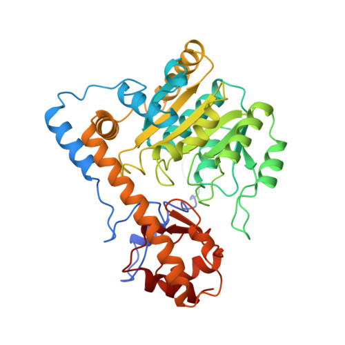
Substitution of apolar residues in the active site of aspartate aminotransferase by histidine. Effects on reaction and substrate specificity.
Vacca, R.A., Christen, P., Malashkevich, V.N., Jansonius, J.N., Sandmeier, E.(1995) Eur J Biochem 227: 481-487
- PubMed: 7851426
- DOI: https://doi.org/10.1111/j.1432-1033.1995.tb20413.x
- Primary Citation of Related Structures:
1ARI - PubMed Abstract:
In an attempt to change the reaction and substrate specificity of aspartate aminotransferase, several apolar active-site residues were substituted in turn with a histidine residue. Aspartate aminotransferase W140H (of Escherichia coli) racemizes alanine seven times faster (Kcat' = 2.2 x 10(-4) s-1) than the wild-type enzyme, while the aminotransferase activity toward L-alanine was sixfold decreased. X-ray crystallographic analysis showed that the structural changes brought about by the mutation are limited to the immediate environment of H140. In contrast to the tryptophan side chain in the wild-type structure, the imidazole ring of H140 does not form a stacking interaction with the coenzyme pyridine ring. The angle between the two ring planes is about 50 degrees. Pyridoxamine 5'-phosphate dissociates 50 times more rapidly from the W140H mutant than from the wild-type enzyme. A model of the structure of the quinonoid enzyme substrate intermediate indicates that H140 might assist in the reprotonation of C alpha of the amino acid substrate from the re side of the deprotonated coenzyme-substrate adduct in competition with si-side reprotonation by K258. In aspartate aminotransferase I17H (of chicken mitochondria), the substituted residue also lies on the re side of the coenzyme. This mutant enzyme slowly decarboxylates L-aspartate to L-alanine (Kcat' = 8 x 10(-5) s-1). No beta-decarboxylase activity is detectable in the wild-type enzyme. In aspartate aminotransferase V37H (of chicken mitochondria), the mutated residue lies besides the coenzyme in the plane of the pyridine ring; no change in reaction specificity was observed. All three mutations, i.e. W140-->H, I17-->H and V37--H, decreased the aminotransferase activity toward aromatic amino acids by 10-100-fold, while decreasing the activity toward dicarboxylic substrates only moderately to 20%, 20% and 60% of the activity of the wild-type enzymes, respectively. In all three mutant enzymes, the decrease in aspartate aminotransferase activity at pH values lower than 6.5 was more pronounced than in the wild-type enzyme, apparently due to the protonation of the newly introduced histidine residues. The study shows that substitutions of single active-site residues may result in altered reaction and substrate specificities of pyridoxal-5'-phosphate-dependent enzymes.
Organizational Affiliation:
Centro di Studio sui Mitocondri e Metabolismo Energetico, Consiglio Nazionale delle Ricerche, Università di Bari, Italy.