1AMH

UNCOMPLEXED RAT TRYPSIN MUTANT WITH ASP 189 REPLACED WITH SER (D189S)


Experimental Data Snapshot

  • Method: X-RAY DIFFRACTION
  • Resolution: 2.50 Å
  • R-Value Free: 0.242 
  • R-Value Work: 0.182 
  • R-Value Observed: 0.182 

wwPDB Validation   3D Report Full Report


This is version 1.4 of the entry. See complete history

Re-refinement Note

A newer entry is available that reflects an alternative modeling of the original data: 8AST 8ASU


Literature

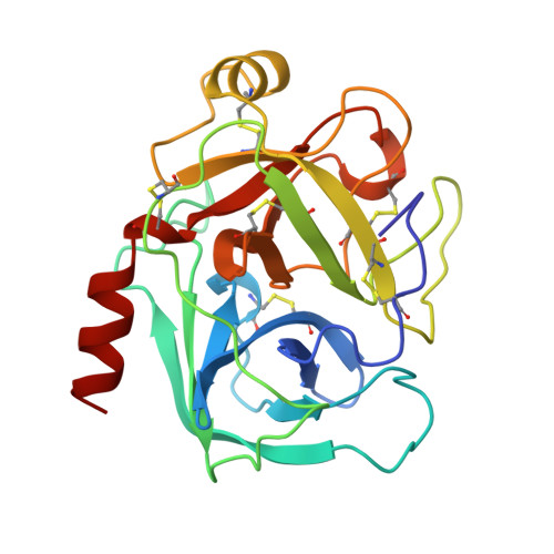
The three-dimensional structure of Asp189Ser trypsin provides evidence for an inherent structural plasticity of the protease.

Szabo, E.Bocskei, Z.Naray-Szabo, G.Graf, L.

(1999) Eur J Biochem 263: 20-26

  • DOI: https://doi.org/10.1046/j.1432-1327.1999.00452.x
  • Primary Citation of Related Structures:  
    1AMH

  • PubMed Abstract: 

    Trypsin mutant Asp189Ser, first described by Gráf et al. [Gráf, L., Jancsó, A., Szilágyi, L., Hegyi, G., Pintér, K., Náray-Szabó, G., Hepp, J., Medzihradszky, K. & Rutter, W.J. (1988) Proc. Natl Acad. Sci. USA 85, 4961-4965] has played an important role in recent studies on the structural basis of substrate-specific catalysis by serine proteases. The present work reports the three-dimensional structure of this mutant crystallized in unliganded form: the first unliganded rat trypsin structure reported. The X-ray structure of the Asp189Ser trypsin mutant in complex with bovine pancreatic trypsin inhibitor is already known. The X-ray structure of free Asp189Ser rat trypsin revealed that the single amino acid mutation at the bottom of the substrate binding pocket of trypsin resulted in extensive structural changes around the mutated site and in dimerization of the mutant, in contrast with the complexed enzyme the structure of which is practically the same as that of wild-type trypsin. The structural rearrangement in the mutant was shown to be restricted to the activation domain region providing further evidence for the allosteric property of this structural-functional unit of the enzyme. This study supports our view that the plasticity of the activation domain may play an important role in the mechanism of substrate-specific serine protease action.


  • Organizational Affiliation

    Department of Biochemistry, Eötvös L. University, Budapest, Hungary.


Macromolecules
Find similar proteins by:  (by identity cutoff)  |  3D Structure
Entity ID: 1
MoleculeChains Sequence LengthOrganismDetailsImage
ANIONIC TRYPSIN
A, B
223Rattus rattusMutation(s): 1 
EC: 3.4.21.4
UniProt
Find proteins for P00763 (Rattus norvegicus)
Explore P00763 
Go to UniProtKB:  P00763
Entity Groups  
Sequence Clusters30% Identity50% Identity70% Identity90% Identity95% Identity100% Identity
UniProt GroupP00763
Sequence Annotations
Expand
  • Reference Sequence
Experimental Data & Validation

Experimental Data

  • Method: X-RAY DIFFRACTION
  • Resolution: 2.50 Å
  • R-Value Free: 0.242 
  • R-Value Work: 0.182 
  • R-Value Observed: 0.182 
  • Space Group: P 1 21 1
Unit Cell:
Length ( Å )Angle ( ˚ )
a = 45.92α = 90
b = 49.79β = 97.34
c = 89.88γ = 90
Software Package:
Software NamePurpose
bioteXdata collection
bioteXdata reduction
X-PLORmodel building
X-PLORrefinement
bioteXdata scaling
X-PLORphasing

Structure Validation

View Full Validation Report


Entry History 

Deposition Data

Revision History  (Full details and data files)

  • Version 1.0: 1997-12-24
    Type: Initial release
  • Version 1.1: 2008-03-04
    Changes: Version format compliance
  • Version 1.2: 2011-07-13
    Changes: Version format compliance
  • Version 1.3: 2021-11-03
    Changes: Database references, Derived calculations, Other, Refinement description
  • Version 1.4: 2023-08-02
    Changes: Refinement description