1ADV

EARLY E2A DNA-BINDING PROTEIN


Experimental Data Snapshot

  • Method: X-RAY DIFFRACTION
  • Resolution: 3.20 Å
  • R-Value Free: 0.336 
  • R-Value Work: 0.206 
  • R-Value Observed: 0.206 

wwPDB Validation   3D Report Full Report


This is version 1.3 of the entry. See complete history


Literature

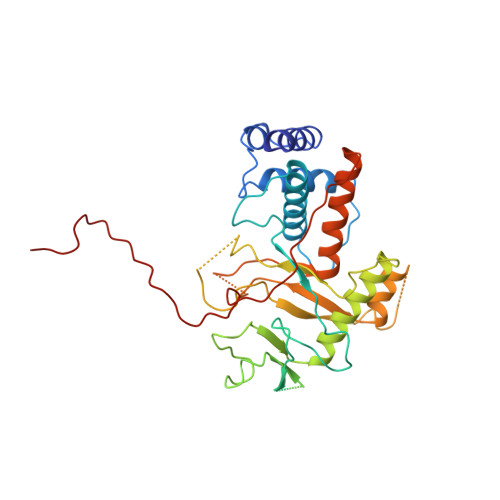
Alternative arrangements of the protein chain are possible for the adenovirus single-stranded DNA binding protein.

Kanellopoulos, P.N.Tsernoglou, D.van der Vliet, P.C.Tucker, P.A.

(1996) J Mol Biol 257: 1-8

  • DOI: https://doi.org/10.1006/jmbi.1996.0141
  • Primary Citation of Related Structures:  
    1ADU, 1ADV

  • PubMed Abstract: 

    A second crystal form of the C-terminal domain of the adenovirus single-stranded DNA binding protein crystallizes in space group P2(1)2(1)2(1) with a=61.0 angstrom, b=91.2 angstrom and c=149.4 angstrom. The crystals contain two molecules per asymmetric unit and diffract to a maximum resolution of 3.0 angstrom. The crystal is composed of infinite chains of molecules along the crystallographic 2(1) axis parallel to c. The principal intermolecular interaction is a hooking of the C-terminal 17 residues of one molecule onto the next molecule in the protein chain. Adjacent molecules in the chain are rotated approximately 90 degrees with respect to their neighbours. The difference in relative orientation of adjacent molecules between the two crystal forms of the protein implies a degree of flexibility in the protein chain that would facilitate DNA binding.


  • Organizational Affiliation

    Structural Biology Programme, European Molecular Biology Laboratory, Heidelberg, Germany.


Macromolecules
Find similar proteins by:  (by identity cutoff)  |  3D Structure
Entity ID: 1
MoleculeChains Sequence LengthOrganismDetailsImage
ADENOVIRUS SINGLE-STRANDED DNA-BINDING PROTEIN
A, B
356Human adenovirus 5Mutation(s): 0 
UniProt
Find proteins for P03265 (Human adenovirus C serotype 5)
Explore P03265 
Go to UniProtKB:  P03265
Entity Groups  
Sequence Clusters30% Identity50% Identity70% Identity90% Identity95% Identity100% Identity
UniProt GroupP03265
Sequence Annotations
Expand
  • Reference Sequence
Experimental Data & Validation

Experimental Data

  • Method: X-RAY DIFFRACTION
  • Resolution: 3.20 Å
  • R-Value Free: 0.336 
  • R-Value Work: 0.206 
  • R-Value Observed: 0.206 
  • Space Group: P 21 21 21
Unit Cell:
Length ( Å )Angle ( ˚ )
a = 61.37α = 90
b = 89.58β = 90
c = 149.77γ = 90
Software Package:
Software NamePurpose
X-PLORmodel building
X-PLORrefinement
XDSdata reduction
DENZOdata reduction
X-PLORphasing

Structure Validation

View Full Validation Report


Entry History 

Deposition Data

Revision History  (Full details and data files)

  • Version 1.0: 1996-06-20
    Type: Initial release
  • Version 1.1: 2008-03-24
    Changes: Version format compliance
  • Version 1.2: 2011-07-13
    Changes: Version format compliance
  • Version 1.3: 2024-02-07
    Changes: Data collection, Database references, Derived calculations, Other, Structure summary