1ADQ

CRYSTAL STRUCTURE OF A HUMAN IGM RHEUMATOID FACTOR FAB IN COMPLEX WITH ITS AUTOANTIGEN IGG FC


Experimental Data Snapshot

  • Method: X-RAY DIFFRACTION
  • Resolution: 3.15 Å
  • R-Value Free: 0.289 
  • R-Value Work: 0.225 
  • R-Value Observed: 0.225 

wwPDB Validation   3D Report Full Report


This is version 1.3 of the entry. See complete history


Literature

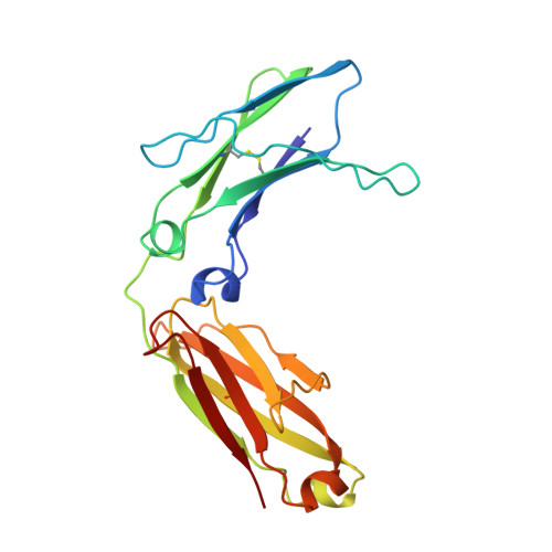
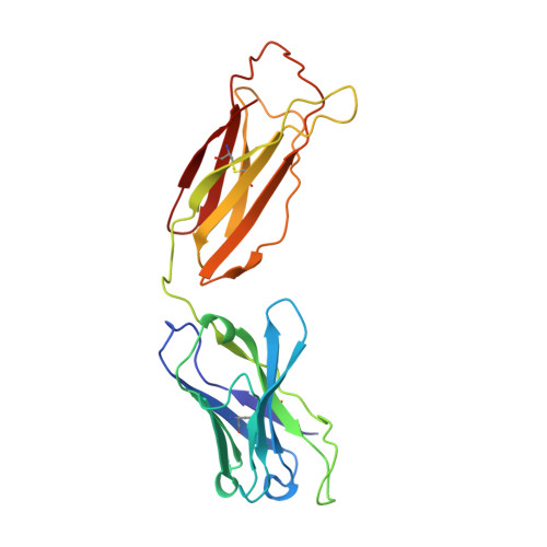
Structure of human IgM rheumatoid factor Fab bound to its autoantigen IgG Fc reveals a novel topology of antibody-antigen interaction.

Corper, A.L.Sohi, M.K.Bonagura, V.R.Steinitz, M.Jefferis, R.Feinstein, A.Beale, D.Taussig, M.J.Sutton, B.J.

(1997) Nat Struct Biol 4: 374-381

  • DOI: https://doi.org/10.1038/nsb0597-374
  • Primary Citation of Related Structures:  
    1ADQ

  • PubMed Abstract: 

    Rheumatoid factors are the characteristic autoantibodies of rheumatoid arthritis, which bind to the Fc regions of IgG molecules. Here we report the crystal structure of the Fab fragment of a patient-derived IgM rheumatoid factor (RF-AN) complexed with human IgG4 Fc, at 3.2 A resolution. This is the first structure of an autoantibody-autoantigen complex. The epitope recognised in IgG Fc includes the C gamma 2/C gamma 3 cleft region, and overlaps the binding sites of bacterial Fc-binding proteins. The antibody residues involved in autorecognition are all located at the edge of the conventional combining site surface, leaving much of the latter available, potentially, for recognition of a different antigen. Since an important contact residue is somatic mutation, the structure implicates antigen-driven selection, following somatic mutation of germline genes, in the production of pathogenic rheumatoid factors.


  • Organizational Affiliation

    Randall Institute, King's College London, UK.


Macromolecules
Find similar proteins by:  (by identity cutoff)  |  3D Structure
Entity ID: 1
MoleculeChains Sequence LengthOrganismDetailsImage
IGG4 REA FC206Homo sapiensMutation(s): 0 
UniProt & NIH Common Fund Data Resources
Find proteins for P01861 (Homo sapiens)
Explore P01861 
Go to UniProtKB:  P01861
PHAROS:  P01861
GTEx:  ENSG00000211892 
Entity Groups  
Sequence Clusters30% Identity50% Identity70% Identity90% Identity95% Identity100% Identity
UniProt GroupP01861
Sequence Annotations
Expand
  • Reference Sequence
Find similar proteins by:  (by identity cutoff)  |  3D Structure
Entity ID: 2
MoleculeChains Sequence LengthOrganismDetailsImage
IGM-LAMBDA RF-AN FAB (LIGHT CHAIN)B [auth L]213Homo sapiensMutation(s): 0 
Entity Groups  
Sequence Clusters30% Identity50% Identity70% Identity90% Identity95% Identity100% Identity
Sequence Annotations
Expand
  • Reference Sequence
Find similar proteins by:  (by identity cutoff)  |  3D Structure
Entity ID: 3
MoleculeChains Sequence LengthOrganismDetailsImage
IGM-LAMBDA RF-AN FAB (HEAVY CHAIN)C [auth H]225Homo sapiensMutation(s): 0 
Entity Groups  
Sequence Clusters30% Identity50% Identity70% Identity90% Identity95% Identity100% Identity
Sequence Annotations
Expand
  • Reference Sequence
Experimental Data & Validation

Experimental Data

  • Method: X-RAY DIFFRACTION
  • Resolution: 3.15 Å
  • R-Value Free: 0.289 
  • R-Value Work: 0.225 
  • R-Value Observed: 0.225 
  • Space Group: C 1 2 1
Unit Cell:
Length ( Å )Angle ( ˚ )
a = 160.35α = 90
b = 81.95β = 98.27
c = 64.24γ = 90
Software Package:
Software NamePurpose
DENZOdata reduction
CCP4data reduction
AMoREphasing
X-PLORrefinement
CCP4data scaling

Structure Validation

View Full Validation Report


Entry History 

Deposition Data

Revision History  (Full details and data files)

  • Version 1.0: 1998-09-16
    Type: Initial release
  • Version 1.1: 2008-03-24
    Changes: Version format compliance
  • Version 1.2: 2011-07-13
    Changes: Version format compliance
  • Version 1.3: 2023-08-02
    Changes: Database references, Refinement description