1A6Z

HFE (HUMAN) HEMOCHROMATOSIS PROTEIN


Experimental Data Snapshot

  • Method: X-RAY DIFFRACTION
  • Resolution: 2.60 Å
  • R-Value Free: 0.277 
  • R-Value Work: 0.233 
  • R-Value Observed: 0.233 

wwPDB Validation   3D Report Full Report


This is version 1.3 of the entry. See complete history


Literature

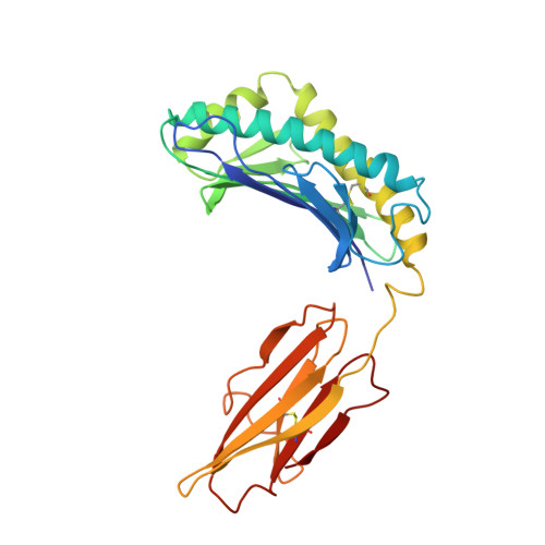
Crystal structure of the hemochromatosis protein HFE and characterization of its interaction with transferrin receptor.

Lebron, J.A.Bennett, M.J.Vaughn, D.E.Chirino, A.J.Snow, P.M.Mintier, G.A.Feder, J.N.Bjorkman, P.J.

(1998) Cell 93: 111-123

  • DOI: https://doi.org/10.1016/s0092-8674(00)81151-4
  • Primary Citation of Related Structures:  
    1A6Z

  • PubMed Abstract: 

    HFE is an MHC-related protein that is mutated in the iron-overload disease hereditary hemochromatosis. HFE binds to transferrin receptor (TfR) and reduces its affinity for iron-loaded transferrin, implicating HFE in iron metabolism. The 2.6 A crystal structure of HFE reveals the locations of hemochromatosis mutations and a patch of histidines that could be involved in pH-dependent interactions. We also demonstrate that soluble TfR and HFE bind tightly at the basic pH of the cell surface, but not at the acidic pH of intracellular vesicles. TfR:HFE stoichiometry (2:1) differs from TfR:transferrin stoichiometry (2:2), implying a different mode of binding for HFE and transferrin to TfR, consistent with our demonstration that HFE, transferrin, and TfR form a ternary complex.


  • Organizational Affiliation

    Division of Biology, California Institute of Technology, Pasadena 91125, USA.


Macromolecules
Find similar proteins by:  (by identity cutoff)  |  3D Structure
Entity ID: 1
MoleculeChains Sequence LengthOrganismDetailsImage
HFE
A, C
275Homo sapiensMutation(s): 0 
Gene Names: HFE
UniProt & NIH Common Fund Data Resources
Find proteins for Q30201 (Homo sapiens)
Explore Q30201 
Go to UniProtKB:  Q30201
PHAROS:  Q30201
GTEx:  ENSG00000010704 
Entity Groups  
Sequence Clusters30% Identity50% Identity70% Identity90% Identity95% Identity100% Identity
UniProt GroupQ30201
Sequence Annotations
Expand
  • Reference Sequence
Find similar proteins by:  (by identity cutoff)  |  3D Structure
Entity ID: 2
MoleculeChains Sequence LengthOrganismDetailsImage
BETA-2-MICROGLOBULIN
B, D
99Homo sapiensMutation(s): 0 
UniProt & NIH Common Fund Data Resources
Find proteins for P61769 (Homo sapiens)
Explore P61769 
Go to UniProtKB:  P61769
PHAROS:  P61769
GTEx:  ENSG00000166710 
Entity Groups  
Sequence Clusters30% Identity50% Identity70% Identity90% Identity95% Identity100% Identity
UniProt GroupP61769
Sequence Annotations
Expand
  • Reference Sequence
Experimental Data & Validation

Experimental Data

  • Method: X-RAY DIFFRACTION
  • Resolution: 2.60 Å
  • R-Value Free: 0.277 
  • R-Value Work: 0.233 
  • R-Value Observed: 0.233 
  • Space Group: P 21 21 21
Unit Cell:
Length ( Å )Angle ( ˚ )
a = 68.8α = 90
b = 100.1β = 90
c = 147.6γ = 90
Software Package:
Software NamePurpose
DENZOdata reduction
SCALEPACKdata scaling
AMoREphasing
X-PLORrefinement

Structure Validation

View Full Validation Report


Entry History 

Deposition Data

Revision History  (Full details and data files)

  • Version 1.0: 1999-03-23
    Type: Initial release
  • Version 1.1: 2008-03-24
    Changes: Version format compliance
  • Version 1.2: 2011-07-13
    Changes: Version format compliance
  • Version 1.3: 2023-08-02
    Changes: Database references, Refinement description