1A15

SDF-1ALPHA


Experimental Data Snapshot

  • Method: X-RAY DIFFRACTION
  • Resolution: 2.20 Å
  • R-Value Free: 0.298 
  • R-Value Work: 0.233 
  • R-Value Observed: 0.233 

wwPDB Validation   3D Report Full Report


This is version 1.3 of the entry. See complete history


Literature

Crystal structure of chemically synthesized [N33A] stromal cell-derived factor 1alpha, a potent ligand for the HIV-1 "fusin" coreceptor.

Dealwis, C.Fernandez, E.J.Thompson, D.A.Simon, R.J.Siani, M.A.Lolis, E.

(1998) Proc Natl Acad Sci U S A 95: 6941-6946

  • DOI: https://doi.org/10.1073/pnas.95.12.6941
  • Primary Citation of Related Structures:  
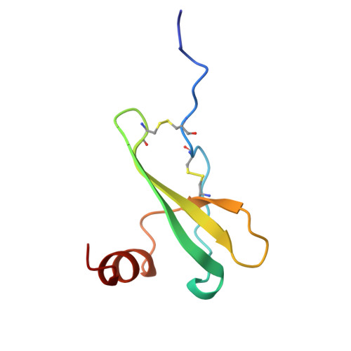
    1A15

  • PubMed Abstract: 

    Stromal cell-derived factor-1alpha (SDF-1alpha ) is a member of the chemokine superfamily and functions as a growth factor and chemoattractant through activation of CXCR4/LESTR/Fusin, a G protein-coupled receptor. This receptor also functions as a coreceptor for T-tropic syncytium-inducing strains of HIV-1. SDF-1alpha antagonizes infectivity of these strains by competing with gp120 for binding to the receptor. The crystal structure of a variant SDF-1alpha ([N33A]SDF-1alpha ) prepared by total chemical synthesis has been refined to 2.2-A resolution. Although SDF-1alpha adopts a typical chemokine beta-beta-beta-alpha topology, the packing of the alpha-helix against the beta-sheet is strikingly different. Comparison of SDF-1alpha with other chemokine structures confirms the hypothesis that SDF-1alpha may be either an ancestral protein from which all other chemokines evolved or the chemokine that is the least divergent from a primordial chemokine. The structure of SDF-1alpha reveals a positively charged surface ideal for binding to the negatively charged extracellular loops of the CXCR4 HIV-1 coreceptor. This ionic complementarity is likely to promote the interaction of the mobile N-terminal segment of SDF-1alpha with interhelical sites of the receptor, resulting in a biological response.


  • Organizational Affiliation

    Department of Pharmacology, Yale University School of Medicine, New Haven CT 06510, USA.


Macromolecules
Find similar proteins by:  (by identity cutoff)  |  3D Structure
Entity ID: 1
MoleculeChains Sequence LengthOrganismDetailsImage
STROMAL DERIVED FACTOR-1ALPHA
A, B
67N/AMutation(s): 1 
UniProt & NIH Common Fund Data Resources
Find proteins for P48061 (Homo sapiens)
Explore P48061 
Go to UniProtKB:  P48061
PHAROS:  P48061
GTEx:  ENSG00000107562 
Entity Groups  
Sequence Clusters30% Identity50% Identity70% Identity90% Identity95% Identity100% Identity
UniProt GroupP48061
Sequence Annotations
Expand
  • Reference Sequence
Small Molecules
Ligands 1 Unique
IDChains Name / Formula / InChI Key2D Diagram3D Interactions
SO4
Query on SO4

Download Ideal Coordinates CCD File 
C [auth A]SULFATE ION
O4 S
QAOWNCQODCNURD-UHFFFAOYSA-L
Experimental Data & Validation

Experimental Data

  • Method: X-RAY DIFFRACTION
  • Resolution: 2.20 Å
  • R-Value Free: 0.298 
  • R-Value Work: 0.233 
  • R-Value Observed: 0.233 
  • Space Group: P 21 21 21
Unit Cell:
Length ( Å )Angle ( ˚ )
a = 38.84α = 90
b = 50.47β = 90
c = 64.72γ = 90
Software Package:
Software NamePurpose
DENZOdata reduction
SCALEPACKdata scaling
X-PLORmodel building
X-PLORrefinement
X-PLORphasing

Structure Validation

View Full Validation Report


Entry History 

Deposition Data

Revision History  (Full details and data files)

  • Version 1.0: 1998-08-12
    Type: Initial release
  • Version 1.1: 2008-03-24
    Changes: Version format compliance
  • Version 1.2: 2011-07-13
    Changes: Version format compliance
  • Version 1.3: 2021-11-03
    Changes: Database references, Derived calculations