4Q28

Crystal Structure of the Plectin 1 and 2 Repeats of the Human Periplakin. Northeast Structural Genomics Consortium (NESG) Target HR9083A


Experimental Data Snapshot

  • Method: X-RAY DIFFRACTION
  • Resolution: 2.64 Å
  • R-Value Free: 0.257 
  • R-Value Work: 0.214 
  • R-Value Observed: 0.216 

wwPDB Validation   3D Report Full Report


This is version 1.0 of the entry. See complete history


Literature

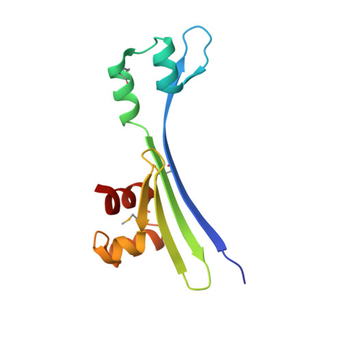
Crystal Structure of the Plectin 1 and 2 Repeats of Human Periplakin.

Vorobiev, S.Lew, S.Seetharaman, J.Janjua, H.Xiao, R.O'Connell, P.T.Maglaqui, M.Wang, D.Everett, J.K.Acton, T.B.Montelione, G.T.Tong, L.Hunt, J.F.

To be published.

Macromolecules
Find similar proteins by:  (by identity cutoff)  |  3D Structure
Entity ID: 1
MoleculeChains Sequence LengthOrganismDetailsImage
Periplakin
A, B, C, D
113Homo sapiensMutation(s): 0 
Gene Names: KIAA0568PPL
UniProt & NIH Common Fund Data Resources
Find proteins for O60437 (Homo sapiens)
Explore O60437 
Go to UniProtKB:  O60437
PHAROS:  O60437
GTEx:  ENSG00000118898 
Entity Groups  
Sequence Clusters30% Identity50% Identity70% Identity90% Identity95% Identity100% Identity
UniProt GroupO60437
Sequence Annotations
Expand
  • Reference Sequence
Small Molecules
Modified Residues  1 Unique
IDChains TypeFormula2D DiagramParent
MSE
Query on MSE
A, B, C, D
L-PEPTIDE LINKINGC5 H11 N O2 SeMET
Experimental Data & Validation

Experimental Data

  • Method: X-RAY DIFFRACTION
  • Resolution: 2.64 Å
  • R-Value Free: 0.257 
  • R-Value Work: 0.214 
  • R-Value Observed: 0.216 
  • Space Group: P 31 2 1
Unit Cell:
Length ( Å )Angle ( ˚ )
a = 68.237α = 90
b = 68.237β = 90
c = 192.587γ = 120
Software Package:
Software NamePurpose
PHENIXrefinement
PDB_EXTRACTdata extraction
HKL-2000data collection
HKL-2000data reduction
HKL-2000data scaling
SHELXSphasing

Structure Validation

View Full Validation Report


Entry History 

Revision History  (Full details and data files)

  • Version 1.0: 2014-06-18
    Type: Initial release