


Crystal Structure of the Q18CP6_CLOD6 protein from glyoxalase family.
Vorobiev, S., Seetharaman, J., Sahdev, S., Xiao, R., Ciccosanti, C., Wang, H., Everett, J.K., Acton, T.B., Montelione, G.T., Tong, L., Hunt, J.F.To be published.
Experimental Data Snapshot
wwPDB Validation 3D Report Full Report

Entity ID: 1 | |||||
|---|---|---|---|---|---|
| Molecule | Chains | Sequence Length | Organism | Details | Image |
| Putative enzyme, glyoxalase family | 163 | Clostridioides difficile 630 | Mutation(s): 0 Gene Names: CD630_01590 | 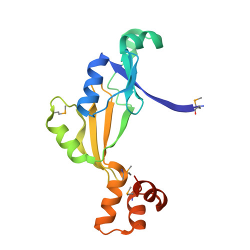 | |
UniProt | |||||
Find proteins for Q18CP6 (Clostridioides difficile (strain 630)) Explore Q18CP6 Go to UniProtKB: Q18CP6 | |||||
Entity Groups | |||||
| Sequence Clusters | 30% Identity50% Identity70% Identity90% Identity95% Identity100% Identity | ||||
| UniProt Group | Q18CP6 | ||||
Sequence AnnotationsExpand | |||||
| |||||
| Modified Residues 1 Unique | |||||
|---|---|---|---|---|---|
| ID | Chains | Type | Formula | 2D Diagram | Parent |
| MSE Query on MSE | A, B | L-PEPTIDE LINKING | C5 H11 N O2 Se |  | MET |
| Length ( Å ) | Angle ( ˚ ) |
|---|---|
| a = 50.388 | α = 90 |
| b = 52.168 | β = 90 |
| c = 135.947 | γ = 90 |
| Software Name | Purpose |
|---|---|
| PHENIX | refinement |
| PDB_EXTRACT | data extraction |
| ADSC | data collection |
| HKL-2000 | data reduction |
| HKL-2000 | data scaling |
| AutoSol | phasing |
RCSB PDB (citation) is hosted by
RCSB PDB is a member of the

